ジャーナル: Proc Natl Acad Sci U S A / 年: 2009 タイトル: 3D structure of the C3bB complex provides insights into the activation and regulation of the complement alternative pathway convertase. 著者: Eva Torreira / Agustín Tortajada / Tamara Montes / Santiago Rodríguez de Córdoba / Oscar Llorca / 要旨: Generation of the alternative pathway C3-convertase, the central amplification enzyme of the complement cascade, initiates by the binding of factor B (fB) to C3b to form the proconvertase, C3bB. C3bB ...Generation of the alternative pathway C3-convertase, the central amplification enzyme of the complement cascade, initiates by the binding of factor B (fB) to C3b to form the proconvertase, C3bB. C3bB is subsequently cleaved by factor D (fD) at a single site in fB, producing Ba and Bb fragments. Ba dissociates from the complex, while Bb remains bound to C3b, forming the active alternative pathway convertase, C3bBb. Using single-particle electron microscopy we have determined the 3-dimensional structures of the C3bB and the C3bBb complexes at approximately 27A resolution. The C3bB structure shows that fB undergoes a dramatic conformational change upon binding to C3b. However, the C3b-bound fB structure was easily interpreted after independently fitting the atomic structures of the isolated Bb and Ba fragments. Interestingly, the divalent cation-binding site in the von Willebrand type A domain in Bb faces the C345C domain of C3b, whereas the serine-protease domain of Bb points outwards. The structure also shows that the Ba fragment interacts with C3b separately from Bb at the level of the alpha'NT and CUB domains. Within this conformation, the long and flexible linker between Bb and Ba is likely exposed and accessible for cleavage by fD to form the active convertase, C3bBb. The architecture of the C3bB and C3bBb complexes reveals that C3b could promote cleavage and activation of fB by actively displacing the Ba domain from the von Willebrand type A domain in free fB. These structures provide a structural basis to understand fundamental aspects of the activation and regulation of the alternative pathway C3-convertase.
試料ホルダー: Eucentric / 試料ホルダーモデル: OTHER / Tilt angle max: 35
撮影
カテゴリ: FILM / フィルム・検出器のモデル: KODAK SO-163 FILM / デジタル化 - スキャナー: OTHER / デジタル化 - サンプリング間隔: 10.5 µm 詳細: images scanned with a MINOLTA Dimage Scan Multi Pro scanner at 2400 dpi and averaged to a final 4.2 angstroms pixel at the specimen ビット/ピクセル: 16
 ムービー
ムービー コントローラー


 データを開く
 基本情報
基本情報 マップデータ
 試料
試料 キーワード
キーワード
 Homo sapiens (ヒト)
 データ登録者
 引用
引用
 構造の表示
構造の表示 ムービービューア
 ダウンロードとリンク
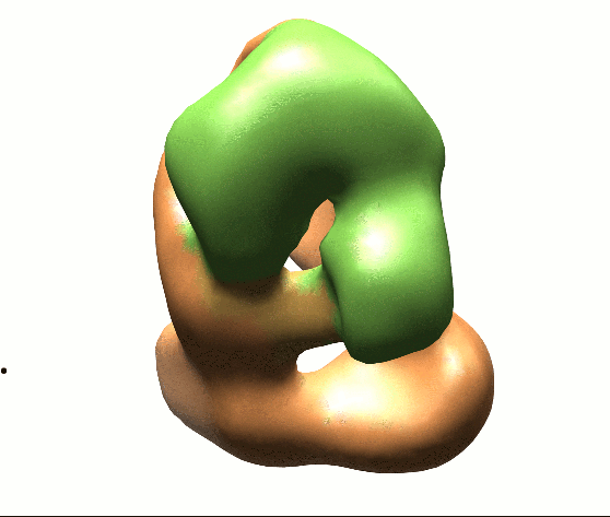 1583.gif
 http://ftp.pdbj.org/pub/emdb/structures/EMD-1583


 Z (Sec.)
 Y (Row.)
 X (Col.)


 試料の構成要素
 解析
解析 電子顕微鏡法
