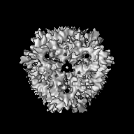
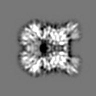
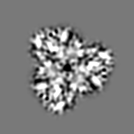
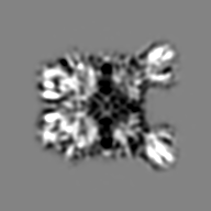
- EMDB-1440: The subnanometer resolution structure of the glutamate synthase 1... -
+
データを開く
IDまたはキーワード:
読み込み中...
-
基本情報
登録情報
データベース: EMDB / ID: EMD-1440
タイトル
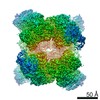
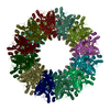
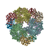
The subnanometer resolution structure of the glutamate synthase 1.2-MDa hexamer by cryoelectron microscopy and its oligomerization behavior in solution: functional implications.
マップデータ
This is the 3D map file of Azospirillum brasilense NADPH-GltS (glutamate synthase)
試料
試料: Azospirillum brasilense NADPH-GltS
タンパク質・ペプチド: NADPH-glutamate synthase
機能・相同性
機能・相同性情報
グルタミン酸シンターゼ (NADPH) / glutamate synthase (NADPH) activity / L-glutamate biosynthetic process / 3 iron, 4 sulfur cluster binding / glutamine metabolic process / 4 iron, 4 sulfur cluster binding / metal ion binding 類似検索 - 分子機能
ジャーナル: J Biol Chem / 年: 2008 タイトル: The subnanometer resolution structure of the glutamate synthase 1.2-MDa hexamer by cryoelectron microscopy and its oligomerization behavior in solution: functional implications. 著者: Magali Cottevieille / Eric Larquet / Slavica Jonic / Maxim V Petoukhov / Gianluca Caprini / Stefano Paravisi / Dmitri I Svergun / Maria A Vanoni / Nicolas Boisset / 要旨: The three-dimensional structure of the hexameric (alphabeta)(6) 1.2-MDa complex formed by glutamate synthase has been determined at subnanometric resolution by combining cryoelectron microscopy, ...The three-dimensional structure of the hexameric (alphabeta)(6) 1.2-MDa complex formed by glutamate synthase has been determined at subnanometric resolution by combining cryoelectron microscopy, small angle x-ray scattering, and molecular modeling, providing for the first time a molecular model of this complex iron-sulfur flavoprotein. In the hexameric species, interprotomeric alpha-alpha and alpha-beta contacts are mediated by the C-terminal domain of the alpha subunit, which is based on a beta helical fold so far unique to glutamate synthases. The alphabeta protomer extracted from the hexameric model is fully consistent with it being the minimal catalytically active form of the enzyme. The structure clarifies the electron transfer pathway from the FAD cofactor on the beta subunit, to the FMN on the alpha subunit, through the low potential [4Fe-4S](1+/2+) centers on the beta subunit and the [3Fe-4S](0/1+) cluster on the alpha subunit. The (alphabeta)(6) hexamer exhibits a concentration-dependent equilibrium with alphabeta monomers and (alphabeta)(2) dimers, in solution, the hexamer being destabilized by high ionic strength and, to a lower extent, by the reaction product NADP(+). Hexamerization seems to decrease the catalytic efficiency of the alphabeta protomer only 3-fold by increasing the K(m) values measured for l-Gln and 2-OG. However, it cannot be ruled out that the (alphabeta)(6) hexamer acts as a scaffold for the assembly of multienzymatic complexes of nitrogen metabolism or that it provides a means to regulate the activity of the enzyme through an as yet unknown ligand.
pH: 7.5 / 詳細: 25 mM Hepes/KOH, 1 mM EDTA, 1 mM DTT
染色
タイプ: NEGATIVE 詳細: CRYOEM : 4 microL were applied on a 200 mesh copper grid, coated with a thin holey carbon film. After blotting the excess of solution with Whatman paper, the grid was rapidly plunged into liquid ethane
グリッド
詳細: 200 mesh copper grid, coated with a thin holey
カテゴリ: FILM / フィルム・検出器のモデル: KODAK SO-163 FILM / デジタル化 - スキャナー: OTHER / デジタル化 - サンプリング間隔: 1.59 µm / 実像数: 151 / 平均電子線量: 10 e/Å2 / 詳細: Scanner model : Nikon Coolscan 8000ED / ビット/ピクセル: 8
Tilt angle min
0
Tilt angle max
0
-
画像解析
CTF補正
詳細: Wiener filtration of volumes from focal series
最終 再構成
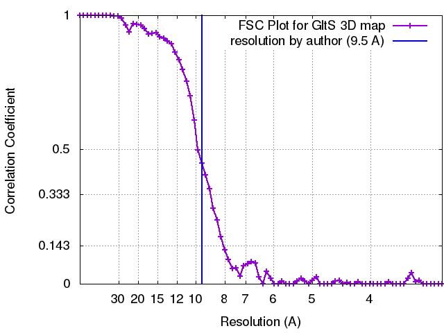
想定した対称性 - 点群: D3 (2回x3回 2面回転対称) アルゴリズム: OTHER / 解像度のタイプ: BY AUTHOR / 解像度: 9.5 Å / 解像度の算出法: FSC 0.5 CUT-OFF / ソフトウェア - 名称: SPIDER / 使用した粒子像数: 12800
詳細
Particles were automatically selected using Roseman algorithm with SPIDER software
FSC曲線 (解像度の算出)
-
原子モデル構築 1
ソフトウェア
名称: Chimera
詳細
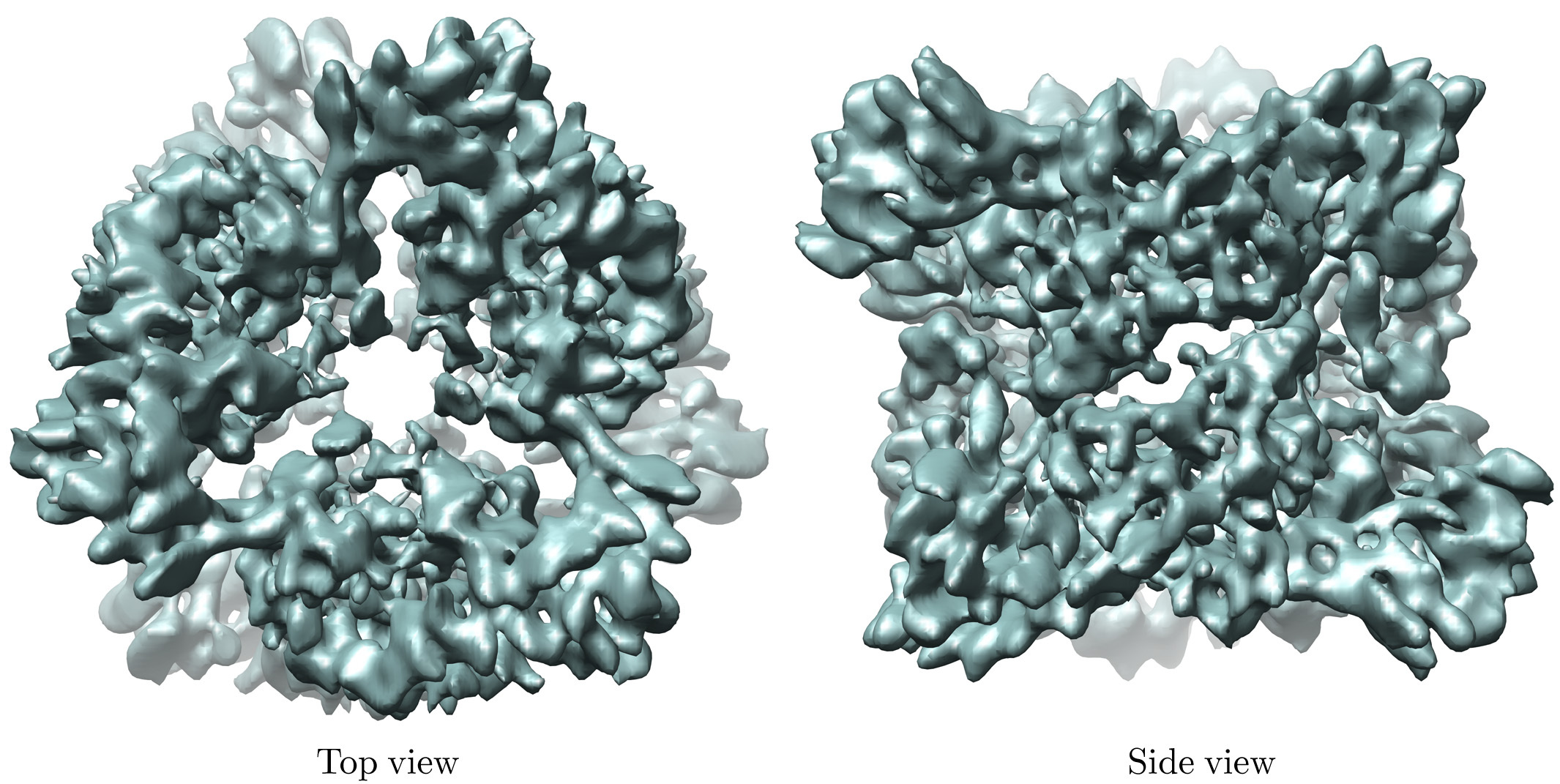
Protocol: Rigid Body. One alpha dimer (1EA0) and one beta model (homology modelling) were separately fitted by manual docking using Chimera software. D3 symmetry was used to reconstruct the whole complex.
精密化
空間: REAL / プロトコル: RIGID BODY FIT
得られたモデル
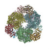
PDB-2vdc: THE 9.5 A RESOLUTION STRUCTURE OF GLUTAMATE SYNTHASE FROM CRYO-ELECTRON MICROSCOPY AND ITS OLIGOMERIZATION BEHAVIOR IN SOLUTION: FUNCTIONAL IMPLICATIONS.
 ムービー
ムービー コントローラー


 データを開く
 基本情報
基本情報 マップデータ
 試料
試料 機能・相同性情報
 グルタミン酸シンターゼ (NADPH) /
 データ登録者
 引用
引用
 構造の表示
構造の表示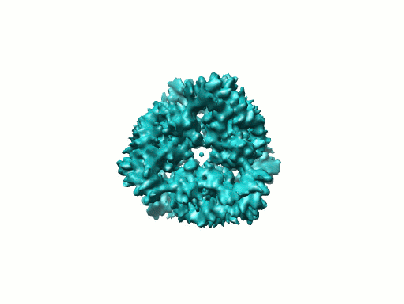

 ダウンロードとリンク
 1440.gif
 http://ftp.pdbj.org/pub/emdb/structures/EMD-1440
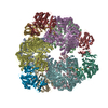


 Z (Sec.)
 Y (Row.)
 X (Col.)


 試料の構成要素
 解析
解析 電子顕微鏡法