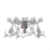
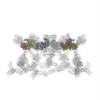
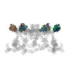
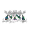
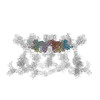
- EMDB-1048: Three-dimensional structure of bacteriophage T4 baseplate. -
+
データを開く
IDまたはキーワード:
読み込み中...
-
基本情報
登録情報
データベース: EMDB / ID: EMD-1048
タイトル
Three-dimensional structure of bacteriophage T4 baseplate.
マップデータ
map contains the baseplate and the proximal part of the tail tube
試料
試料: Bacteriophage T4 baseplate-tail tube assembly
タンパク質・ペプチド: baseplate-tail tube complex
機能・相同性
機能・相同性情報
symbiont entry into host cell via disruption of host cell wall peptidoglycan / virus tail, baseplate / viral tail assembly / virus tail, fiber / symbiont entry into host cell via disruption of host cell envelope / virus tail / symbiont entry into host / viral release from host cell / peptidoglycan catabolic process / cell wall macromolecule catabolic process ...symbiont entry into host cell via disruption of host cell wall peptidoglycan / virus tail, baseplate / viral tail assembly / virus tail, fiber / symbiont entry into host cell via disruption of host cell envelope / virus tail / symbiont entry into host / viral release from host cell / peptidoglycan catabolic process / cell wall macromolecule catabolic process / リゾチーム / lysozyme activity / killing of cells of another organism / entry receptor-mediated virion attachment to host cell / defense response to bacterium / symbiont entry into host cell / identical protein binding / metal ion binding 類似検索 - 分子機能
Bacteriophage T4, Gp12 / Short tail fibre protein, C-terminal / Short tail fibre protein, C-terminal superfamily / Phage short tail fibre protein gp12, middle domain / Short tail fibre protein receptor-binding domain / Phage tail collar domain / Bacteriophage T4, Gp27, baseplate hub, C-terminal / Bacteriophage T4, Gp27, baseplate hub, N-terminal / Phage tail collar domain superfamily / Gp27, baseplate hub, domain 3 ...Bacteriophage T4, Gp12 / Short tail fibre protein, C-terminal / Short tail fibre protein, C-terminal superfamily / Phage short tail fibre protein gp12, middle domain / Short tail fibre protein receptor-binding domain / Phage tail collar domain / Bacteriophage T4, Gp27, baseplate hub, C-terminal / Bacteriophage T4, Gp27, baseplate hub, N-terminal / Phage tail collar domain superfamily / Gp27, baseplate hub, domain 3 / Gp27, baseplate hub, domain 4 / Gp27, baseplate hub, domain 2 / Phage Tail Collar Domain / Baseplate structural protein, domain 2 / Baseplate structural protein, domain 1 / Baseplate structural protein Gp11 / Bacteriophage T4, Gp11, C-terminal finger domain / Baseplate structural protein Gp11, N-terminal domain superfamily / Baseplate structural protein Gp11 superfamily / Baseplate structural protein Gp11, C-terminal domain / GP11 baseplate wedge protein / Baseplate wedge protein gp10 / Baseplate wedge protein gp6 / : / : / : / : / Baseplate wedge protein gp6-like, helical domain / Baseplate structural protein gp6, C-terminal domain I / Baseplate structural protein gp6, C-terminal domain II / Baseplate wedge protein gp6, domain II / Baseplate structural protein Gp9 C-terminal domain superfamily / Bacteriophage T4, Gp8 / Bacteriophage T4, Gp8 superfamily / Bacteriophage T4, Gp8 / Baseplate structural protein Gp9/Gp10 / Baseplate structural protein Gp9/Gp10 middle domain superfamily / Gp9-like superfamily / Bacteriophage T4 gp9/10-like protein / Protein Gp5, N-terminal OB-fold domain / Gp5, C-terminal / Pre-baseplate central spike protein Gp5 / Gp5 N-terminal OB domain / Gp5 C-terminal repeat (3 copies) / T4-type lysozyme / Glycoside hydrolase, family 24 / Lysozyme domain superfamily / Phage lysozyme / Lysozyme-like domain superfamily / Peptidase S1, PA clan, chymotrypsin-like fold 類似検索 - ドメイン・相同性
Baseplate protein gp9 / Baseplate wedge protein gp10 / Baseplate wedge protein gp11 / Short tail fiber protein gp12 / Pre-baseplate central spike protein Gp5 / Baseplate central spike complex protein gp27 / Baseplate wedge protein gp6 / Baseplate wedge protein gp8 類似検索 - 構成要素
ジャーナル: Nat Struct Biol / 年: 2003 タイトル: Three-dimensional structure of bacteriophage T4 baseplate. 著者: Victor A Kostyuchenko / Petr G Leiman / Paul R Chipman / Shuji Kanamaru / Mark J van Raaij / Fumio Arisaka / Vadim V Mesyanzhinov / Michael G Rossmann / 要旨: The baseplate of bacteriophage T4 is a multiprotein molecular machine that controls host cell recognition, attachment, tail sheath contraction and viral DNA ejection. We report here the three- ...The baseplate of bacteriophage T4 is a multiprotein molecular machine that controls host cell recognition, attachment, tail sheath contraction and viral DNA ejection. We report here the three-dimensional structure of the baseplate-tail tube complex determined to a resolution of 12 A by cryoelectron microscopy. The baseplate has a six-fold symmetric, dome-like structure approximately 520 A in diameter and approximately 270 A long, assembled around a central hub. A 940 A-long and 96 A-diameter tail tube, coaxial with the hub, is connected to the top of the baseplate. At the center of the dome is a needle-like structure that was previously identified as a cell puncturing device. We have identified the locations of six proteins with known atomic structures, and established the position and shape of several other baseplate proteins. The baseplate structure suggests a mechanism of baseplate triggering and structural transition during the initial stages of T4 infection.
試料ホルダー: 626 Single Tilt Cryotransfer System / 試料ホルダーモデル: GATAN LIQUID NITROGEN
日付
2001年1月30日
撮影
カテゴリ: FILM / フィルム・検出器のモデル: KODAK SO-163 FILM / デジタル化 - スキャナー: ZEISS SCAI / デジタル化 - サンプリング間隔: 14 µm / 実像数: 15 / 平均電子線量: 25 e/Å2 詳細: images were scanned at 7 micron per pixel and averaged 2x2 to give 14 micron per pixel ビット/ピクセル: 8
Tilt angle min
0
Tilt angle max
0
+
画像解析
CTF補正
詳細: each particle
最終 再構成
想定した対称性 - 点群: C6 (6回回転対称) / アルゴリズム: OTHER / 解像度のタイプ: BY AUTHOR / 解像度: 12.0 Å / 解像度の算出法: FSC 0.5 CUT-OFF / ソフトウェア - 名称: SPIDER 詳細: modified SPIDER version was used to allow reconstruction of the whole baseplate-tail tube assembly 使用した粒子像数: 945
+
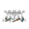
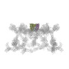
原子モデル構築 1
ソフトウェア
名称: Situs 2.0, Colores
詳細
Protocol: laplacian filtered real space
精密化
空間: REAL / プロトコル: RIGID BODY FIT / 当てはまり具合の基準: correlation coefficient
得られたモデル
PDB-1pdf: Fitting of gp11 crystal structure into 3D cryo-EM reconstruction of bacteriophage T4 baseplate-tail tube complex
PDB-1pdi: Fitting of the C-terminal part of the short tail fibers into the cryo-EM reconstruction of T4 baseplate
PDB-1pdj: Fitting of gp27 into cryoEM reconstruction of bacteriophage T4 baseplate
PDB-1pdl: Fitting of gp5 in the cryoEM reconstruction of the bacteriophage T4 baseplate
PDB-1pdm: Fitting of gp8 structure into the cryoEM reconstruction of the bacteriophage T4 baseplate
PDB-1pdp: Fitting of gp9 structure into the bacteriophage T4 baseplate cryoEM reconstruction
PDB-2fl8: Fitting of the gp10 trimer structure into the cryoEM map of the bacteriophage T4 baseplate in the hexagonal conformation.
PDB-3h3w: Fitting of the gp6 crystal structure into 3D cryo-EM reconstruction of bacteriophage T4 dome-shaped baseplate
+
原子モデル構築 2
ソフトウェア
名称: Situs 2.0, Colores
詳細
Protocol: laplacian filtered real space
精密化
空間: REAL / プロトコル: RIGID BODY FIT / 当てはまり具合の基準: correlation coefficient
得られたモデル
PDB-1pdf: Fitting of gp11 crystal structure into 3D cryo-EM reconstruction of bacteriophage T4 baseplate-tail tube complex
PDB-1pdi: Fitting of the C-terminal part of the short tail fibers into the cryo-EM reconstruction of T4 baseplate
PDB-1pdj: Fitting of gp27 into cryoEM reconstruction of bacteriophage T4 baseplate
PDB-1pdl: Fitting of gp5 in the cryoEM reconstruction of the bacteriophage T4 baseplate
PDB-1pdm: Fitting of gp8 structure into the cryoEM reconstruction of the bacteriophage T4 baseplate
PDB-1pdp: Fitting of gp9 structure into the bacteriophage T4 baseplate cryoEM reconstruction
PDB-2fl8: Fitting of the gp10 trimer structure into the cryoEM map of the bacteriophage T4 baseplate in the hexagonal conformation.
PDB-3h3w: Fitting of the gp6 crystal structure into 3D cryo-EM reconstruction of bacteriophage T4 dome-shaped baseplate
+
原子モデル構築 3
ソフトウェア
名称: Situs 2.0, Colores
詳細
Protocol: laplacian filtered real space
精密化
空間: REAL / プロトコル: RIGID BODY FIT / 当てはまり具合の基準: correlation coefficient
得られたモデル
PDB-1pdf: Fitting of gp11 crystal structure into 3D cryo-EM reconstruction of bacteriophage T4 baseplate-tail tube complex
PDB-1pdi: Fitting of the C-terminal part of the short tail fibers into the cryo-EM reconstruction of T4 baseplate
PDB-1pdj: Fitting of gp27 into cryoEM reconstruction of bacteriophage T4 baseplate
PDB-1pdl: Fitting of gp5 in the cryoEM reconstruction of the bacteriophage T4 baseplate
PDB-1pdm: Fitting of gp8 structure into the cryoEM reconstruction of the bacteriophage T4 baseplate
PDB-1pdp: Fitting of gp9 structure into the bacteriophage T4 baseplate cryoEM reconstruction
PDB-2fl8: Fitting of the gp10 trimer structure into the cryoEM map of the bacteriophage T4 baseplate in the hexagonal conformation.
PDB-3h3w: Fitting of the gp6 crystal structure into 3D cryo-EM reconstruction of bacteriophage T4 dome-shaped baseplate
+
原子モデル構築 4
ソフトウェア
名称: Situs 2.0, Colores
詳細
Protocol: laplacian filtered real space
精密化
空間: REAL / プロトコル: RIGID BODY FIT / 当てはまり具合の基準: correlation coefficient
得られたモデル
PDB-1pdf: Fitting of gp11 crystal structure into 3D cryo-EM reconstruction of bacteriophage T4 baseplate-tail tube complex
PDB-1pdi: Fitting of the C-terminal part of the short tail fibers into the cryo-EM reconstruction of T4 baseplate
PDB-1pdj: Fitting of gp27 into cryoEM reconstruction of bacteriophage T4 baseplate
PDB-1pdl: Fitting of gp5 in the cryoEM reconstruction of the bacteriophage T4 baseplate
PDB-1pdm: Fitting of gp8 structure into the cryoEM reconstruction of the bacteriophage T4 baseplate
PDB-1pdp: Fitting of gp9 structure into the bacteriophage T4 baseplate cryoEM reconstruction
PDB-2fl8: Fitting of the gp10 trimer structure into the cryoEM map of the bacteriophage T4 baseplate in the hexagonal conformation.
PDB-3h3w: Fitting of the gp6 crystal structure into 3D cryo-EM reconstruction of bacteriophage T4 dome-shaped baseplate
+
原子モデル構築 5
ソフトウェア
名称: Situs 2.0, Colores
詳細
Protocol: laplacian filtered real space
精密化
空間: REAL / プロトコル: RIGID BODY FIT / 当てはまり具合の基準: correlation coefficient
得られたモデル
PDB-1pdf: Fitting of gp11 crystal structure into 3D cryo-EM reconstruction of bacteriophage T4 baseplate-tail tube complex
PDB-1pdi: Fitting of the C-terminal part of the short tail fibers into the cryo-EM reconstruction of T4 baseplate
PDB-1pdj: Fitting of gp27 into cryoEM reconstruction of bacteriophage T4 baseplate
PDB-1pdl: Fitting of gp5 in the cryoEM reconstruction of the bacteriophage T4 baseplate
PDB-1pdm: Fitting of gp8 structure into the cryoEM reconstruction of the bacteriophage T4 baseplate
PDB-1pdp: Fitting of gp9 structure into the bacteriophage T4 baseplate cryoEM reconstruction
PDB-2fl8: Fitting of the gp10 trimer structure into the cryoEM map of the bacteriophage T4 baseplate in the hexagonal conformation.
PDB-3h3w: Fitting of the gp6 crystal structure into 3D cryo-EM reconstruction of bacteriophage T4 dome-shaped baseplate
+
原子モデル構築 6
ソフトウェア
名称: Situs 2.0, Colores
詳細
Protocol: laplacian filtered real space
精密化
空間: REAL / プロトコル: RIGID BODY FIT / 当てはまり具合の基準: correlation coefficient
得られたモデル
PDB-1pdf: Fitting of gp11 crystal structure into 3D cryo-EM reconstruction of bacteriophage T4 baseplate-tail tube complex
PDB-1pdi: Fitting of the C-terminal part of the short tail fibers into the cryo-EM reconstruction of T4 baseplate
PDB-1pdj: Fitting of gp27 into cryoEM reconstruction of bacteriophage T4 baseplate
PDB-1pdl: Fitting of gp5 in the cryoEM reconstruction of the bacteriophage T4 baseplate
PDB-1pdm: Fitting of gp8 structure into the cryoEM reconstruction of the bacteriophage T4 baseplate
PDB-1pdp: Fitting of gp9 structure into the bacteriophage T4 baseplate cryoEM reconstruction
PDB-2fl8: Fitting of the gp10 trimer structure into the cryoEM map of the bacteriophage T4 baseplate in the hexagonal conformation.
PDB-3h3w: Fitting of the gp6 crystal structure into 3D cryo-EM reconstruction of bacteriophage T4 dome-shaped baseplate
+
原子モデル構築 7
ソフトウェア
名称: Situs 2.0, Colores
詳細
Protocol: laplacian filtered real space
精密化
空間: REAL / プロトコル: RIGID BODY FIT / 当てはまり具合の基準: correlation coefficient
得られたモデル
PDB-1pdf: Fitting of gp11 crystal structure into 3D cryo-EM reconstruction of bacteriophage T4 baseplate-tail tube complex
PDB-1pdi: Fitting of the C-terminal part of the short tail fibers into the cryo-EM reconstruction of T4 baseplate
PDB-1pdj: Fitting of gp27 into cryoEM reconstruction of bacteriophage T4 baseplate
PDB-1pdl: Fitting of gp5 in the cryoEM reconstruction of the bacteriophage T4 baseplate
PDB-1pdm: Fitting of gp8 structure into the cryoEM reconstruction of the bacteriophage T4 baseplate
PDB-1pdp: Fitting of gp9 structure into the bacteriophage T4 baseplate cryoEM reconstruction
PDB-2fl8: Fitting of the gp10 trimer structure into the cryoEM map of the bacteriophage T4 baseplate in the hexagonal conformation.
PDB-3h3w: Fitting of the gp6 crystal structure into 3D cryo-EM reconstruction of bacteriophage T4 dome-shaped baseplate
+
原子モデル構築 8
ソフトウェア
名称: Situs 2.0, Colores
詳細
Protocol: laplacian filtered real space
精密化
空間: REAL / プロトコル: RIGID BODY FIT / 当てはまり具合の基準: correlation coefficient
得られたモデル
PDB-1pdf: Fitting of gp11 crystal structure into 3D cryo-EM reconstruction of bacteriophage T4 baseplate-tail tube complex
PDB-1pdi: Fitting of the C-terminal part of the short tail fibers into the cryo-EM reconstruction of T4 baseplate
PDB-1pdj: Fitting of gp27 into cryoEM reconstruction of bacteriophage T4 baseplate
PDB-1pdl: Fitting of gp5 in the cryoEM reconstruction of the bacteriophage T4 baseplate
PDB-1pdm: Fitting of gp8 structure into the cryoEM reconstruction of the bacteriophage T4 baseplate
PDB-1pdp: Fitting of gp9 structure into the bacteriophage T4 baseplate cryoEM reconstruction
PDB-2fl8: Fitting of the gp10 trimer structure into the cryoEM map of the bacteriophage T4 baseplate in the hexagonal conformation.
PDB-3h3w: Fitting of the gp6 crystal structure into 3D cryo-EM reconstruction of bacteriophage T4 dome-shaped baseplate
 ムービー
ムービー コントローラー


 データを開く
 基本情報
基本情報 マップデータ
 試料
試料 機能・相同性情報
 リゾチーム /
 データ登録者
 引用
引用
 構造の表示
構造の表示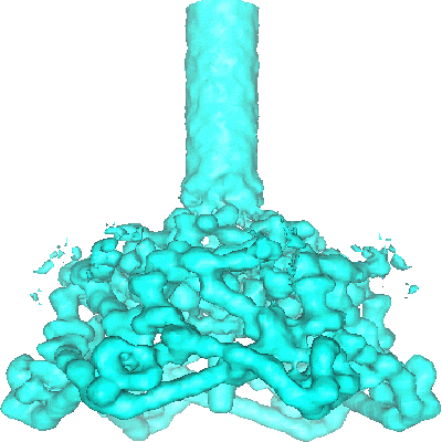
 ダウンロードとリンク
 1048.gif
 http://ftp.pdbj.org/pub/emdb/structures/EMD-1048


 試料の構成要素
 解析
解析 電子顕微鏡法