ジャーナル: J Mol Biol / 年: 2000 タイトル: A new look at the microtubule binding patterns of dimeric kinesins. 著者: A Hoenger / M Thormählen / R Diaz-Avalos / M Doerhoefer / K N Goldie / J Müller / E Mandelkow / 要旨: The interactions of monomeric and dimeric kinesin and ncd constructs with microtubules have been investigated using cryo-electron microscopy (cryo-EM) and several biochemical methods. There is a good ...The interactions of monomeric and dimeric kinesin and ncd constructs with microtubules have been investigated using cryo-electron microscopy (cryo-EM) and several biochemical methods. There is a good consensus on the structure of dimeric ncd when bound to a tubulin dimer showing one head attached directly to tubulin, and the second head tethered to the first. However, the 3D maps of dimeric kinesin motor domains are still quite controversial and leave room for different interpretations. Here we reinvestigated the microtubule binding patterns of dimeric kinesins by cryo-EM and digital 3D reconstruction under different nucleotide conditions and different motor:tubulin ratios, and determined the molecular mass of motor-tubulin complexes by STEM. Both methods revealed complementary results. We found that the ratio of bound kinesin motor-heads to alphabeta-tubulin dimers was never reaching above 1.5 irrespective of the initial mixing ratios. It appears that each kinesin dimer occupies two microtubule-binding sites, provided that there is a free one nearby. Thus the appearances of different image reconstructions can be explained by non-specific excess binding of motor heads. Consequently, the use of different apparent density distributions for docking the X-ray structures onto the microtubule surface leads to different and mutually exclusive models. We propose that in conditions of stoichiometric binding the two heads of a kinesin dimer separate and bind to different tubulin subunits. This is in contrast to ncd where the two heads remain tightly attached on the microtubule surface. Using dimeric kinesin molecules crosslinked in their neck domain we also found that they stabilize protofilaments axially, but not laterally, which is a strong indication that the two heads of the dimers bind along one protofilament, rather than laterally bridging two protofilaments. A molecular walking model based on these results summarizes our conclusions and illustrates the implications of symmetry for such models.
 ムービー
ムービー コントローラー


 データを開く
 基本情報
基本情報 マップデータ
 試料
試料
 Neurospora crassa (アカパンカビ)
 データ登録者
 引用
引用
 構造の表示
構造の表示 ムービービューア
 ダウンロードとリンク
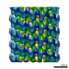
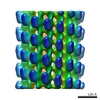
 1035.gif
 http://ftp.pdbj.org/pub/emdb/structures/EMD-1035
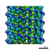
 試料の構成要素
 解析
解析 電子顕微鏡法