 Movie
Movie Controller
[English] 日本語
 Yorodumi
- SASDGY4: Beta-amylase 2, chloroplastic (AtBAM2) (Beta-amylase 2, chloropla... -
+ Open data

- Basic information
| Entry | Database: SASBDB / ID: SASDGY4 |
|---|---|
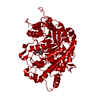
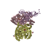
 Sample Sample | Beta-amylase 2, chloroplastic (AtBAM2)
|
| Function / homology |  Function and homology informationbeta-amylase / beta-amylase activity / amylopectin maltohydrolase activity / chloroplast stroma / polysaccharide catabolic process / chloroplast Function and homology informationbeta-amylase / beta-amylase activity / amylopectin maltohydrolase activity / chloroplast stroma / polysaccharide catabolic process / chloroplastSimilarity search - Function |
| Biological species |  Arabidopsis thaliana (thale cress) Arabidopsis thaliana (thale cress) |
 Citation Citation | Date: 2019 Aug 30 Title: Solution structure and assembly of β-amylase 2 from Arabidopsis thaliana Authors: Chandrasekharan N / Ravenburg C / Roy I / Monroe J |
 Contact author Contact author |
|
- Structure visualization
| Structure viewer | Molecule: MolmilJmol/JSmol |
|---|
- Downloads & links
 SASDGY4
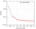

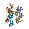
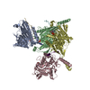
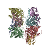
-Models
| Model #3698 |   Type: atomic / Chi-square value: 8.96285214274195  Search similar-shape structures of this assembly by Omokage search (details) Search similar-shape structures of this assembly by Omokage search (details) |
|---|---|
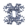
| Model #3699 |   Type: dummy / Symmetry : P222 / Chi-square value: 1.311Search similar-shape structures of this assembly by Omokage search (details) |
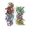
| Model #3700 |   Type: dummy / Software: (dammif (r10552)) / Chi-square value: 2.189 Search similar-shape structures of this assembly by Omokage search (details) |
-Sample
| Sample | Name: Beta-amylase 2, chloroplastic (AtBAM2) / Specimen concentration: 1.65-2.75 |
|---|---|
| Buffer | Name: 50 mM HEPES / pH: 7.5 |
| Entity #1874 | Name: BAM2 / Type: protein / Description: Beta-amylase 2, chloroplastic / Formula weight: 57.141 / Num. of mol.: 4 / Source: Arabidopsis thaliana / References: UniProt: O65258 Sequence: MGSSHHHHHH SQDPAESTEE DRVPIDDDDD STDQLVDEEI VHFEERDFAG TACVPVYVML PLGVIDMNSE VVEPEELLDQ LRTLKSVNVD GVMVDCWWGI VESHTPQVYN WSGYKKLFQM IRELGLKIQV VMSFHECGGN VGDDVHIQIP EWVREIGQSN PDIYFTDSAG ...Sequence: MGSSHHHHHH SQDPAESTEE DRVPIDDDDD STDQLVDEEI VHFEERDFAG TACVPVYVML PLGVIDMNSE VVEPEELLDQ LRTLKSVNVD GVMVDCWWGI VESHTPQVYN WSGYKKLFQM IRELGLKIQV VMSFHECGGN VGDDVHIQIP EWVREIGQSN PDIYFTDSAG RRNTECLTWG IDKQRVLRGR TALEVYFDYM RSFRVEFDEF FEEKIIPEIE VGLGPCGELR YPSYPAQFGW KYPGIGEFQC YDKYLMNSLK EAAEVRGHSF WGRGPDNTET YNSTPHGTGF FRDGGDYDSY YGRFFLNWYS RVLIDHGDRV LAMANLAFEG TCIAAKLSGI HWWYKTASHA AELTAGFYNS SNRDGYGPIA AMFKKHDAAL NFTCVELRTL DQHEDFPEAL ADPEGLVWQV LNAAWDASIP VASENALPCY DREGYNKILE NAKPLTDPDG RHLSCFTYLR LNPTLMESQN FKEFERFLKR MHGEAVPDLG LAPGTQETNP E |
-Experimental information
| Beam | Instrument name: Advanced Light Source (ALS) 12.3.1 (SIBYLS) City: Berkeley, CA / 国: USA  / Type of source: X-ray synchrotronSynchrotron / Wavelength: 0.127 Å / Dist. spec. to detc.: 2 mm / Type of source: X-ray synchrotronSynchrotron / Wavelength: 0.127 Å / Dist. spec. to detc.: 2 mm | |||||||||||||||||||||||||||||||||
|---|---|---|---|---|---|---|---|---|---|---|---|---|---|---|---|---|---|---|---|---|---|---|---|---|---|---|---|---|---|---|---|---|---|---|
| Detector | Name: Pilatus3 X 2M / Pixsize x: 172 mm | |||||||||||||||||||||||||||||||||
| Scan | 
| |||||||||||||||||||||||||||||||||
| Distance distribution function P(R) | 
| |||||||||||||||||||||||||||||||||
| Result |  Comments: We expressed Arabidopsis thaliana AtBAM2 in E. coli BL21 cells from a pETDuet-1 expression vector with an N-terminal 6-His tag as constructed by (Monroe et al., 2017, 2018). We induced ...Comments: We expressed Arabidopsis thaliana AtBAM2 in E. coli BL21 cells from a pETDuet-1 expression vector with an N-terminal 6-His tag as constructed by (Monroe et al., 2017, 2018). We induced AtBAM2 expression in BL21 E coli at OD600 with 0.3 mM IPTG in 2xYT broth with 60 µg/ml ampicillin at 30 oC overnight. Cells were lysed and sonicated in a buffer containing 50 mM NaH2PO4, pH 8, 500 mM NaCl, and 2 mM imidazole. The supernatant was loaded onto a TALON cobalt column using an AKTA Start and washed with a buffer containing 50 mM HEPES pH 8, 500 mM NaCl, 5% glycerol, 10 mM TCEP, and 40 mM imidazole. The protein was then eluted with buffer containing 50 mM HEPES pH 8, 500 mM NaCl, 5% glycerol, 10 mM TCEP, and 500 mM imidazole. Pure protein, as determined by a band present at ∼50 kDa on a 4–20% Tris‐Glycine gel stained with Coomassie Blue, was concentrated in a Spin‐X UF concentrator with a 5000 MWCO. The concentrated protein was then further purified using a HiLoad 16/60 column filled with Superdex 200 in 50 mM HEPES, pH 7. Pure protein based on SDS-PAGE was concentrated as before and the concentration of protein was determined via absorbance at 280 nm using an extinction coefficient of 94310 M−1 cm−1 which was calculated from the sequence using ProtParam (Gasteiger et al., 2005) Matching buffer exposures were collected before and after samples to ensure there was no difference in the scattering due to contamination of the sample cell. Scattering data were subtracted from buffer and then processed in PRIMUS (ATSAS 2.8.4 r10553) to create an average data file (Konarev et al., 2003). Data were analyzed using PRIMUS, GNOM, and SCÅTTER (v3.0g) to determine dimensions and create a merged data file from the two data sets at 30 and 50 µM AtBAM2 (Konarev et al., 2003). We then used the merged data file in DAMMIF (v1.1.2) and DAMMIN (v5.3) to generate the dummy-atom model and aligned the result to the all-atom structure using SASTBX (Svergun, 1992, 1999; Svergun et al., 2001; Franke & Svergun, 2009; Liu et al., 2012). Dummy atom models from different methods were aligned and visualized using YASARA Structure (Krieger et al., 2009).
|
