 Movie
Movie Controller
[English] 日本語
 Yorodumi
- SASDD32: DNA-(adenine N6)-methyltransferase from Acinetobacter baumannii A... -
+ Open data

- Basic information
| Entry | Database: SASBDB / ID: SASDD32 |
|---|---|
 Sample Sample | DNA-(adenine N6)-methyltransferase from Acinetobacter baumannii ATCC 17978
|
| Biological species |   Acinetobacter baumannii ATCC 17978 (bacteria) Acinetobacter baumannii ATCC 17978 (bacteria) |
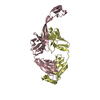
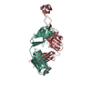
 Citation Citation | Journal: Protein Expr Purif / Year: 2018 Title: Recombinant production of A1S_0222 from Acinetobacter baumannii ATCC 17978 and confirmation of its DNA-(adenine N6)-methyltransferase activity. Authors: Ulrike Blaschke / Beneditta Suwono / Sachli Zafari / Ingo Ebersberger / Evelyn Skiebe / Cy M Jeffries / Dmitri I Svergun / Gottfried Wilharm /  Abstract: Acinetobacter baumannii appears as an often multidrug-resistant nosocomial pathogen in hospitals worldwide. Its remarkable persistence in the hospital environment is probably due to intrinsic and ...Acinetobacter baumannii appears as an often multidrug-resistant nosocomial pathogen in hospitals worldwide. Its remarkable persistence in the hospital environment is probably due to intrinsic and acquired resistance to disinfectants and antibiotics, tolerance to desiccation stress, capability to form biofilms, and is possibly facilitated by surface-associated motility. Our attempts to elucidate surface-associated motility in A. baumannii revealed a mutant inactivated in a putative DNA-(adenine N6)-methyltransferase, designated A1S_0222 in strain ATCC 17978. We recombinantly produced A1S_0222 as a glutathione S-transferase (GST) fusion protein and purified it to near homogeneity through a combination of GST affinity chromatography, cation exchange chromatography and PD-10 desalting column. Furthermore we demonstrate A1S_0222-dependent adenine methylation at a GAATTC site. We propose the name AamA (Acinetobacteradenine methyltransferase A) in addition to the formal names M.AbaBGORF222P/M.Aba17978ORF8565P. Small angle X-ray scattering (SAXS) revealed that the protein is monomeric and has an extended and likely two-domain shape in solution. |
 Contact author Contact author |
|
- Structure visualization
| Structure viewer | Molecule:  MolmilJmol/JSmol MolmilJmol/JSmol |
|---|
- Downloads & links
 SASDD32
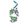


-Models
| Model #1703 |   Type: dummy / Radius of dummy atoms: 2.75 A / Symmetry : P1 / Chi-square value: 1.062 / P-value: 0.132008 Search similar-shape structures of this assembly by Omokage search (details) Search similar-shape structures of this assembly by Omokage search (details) |
|---|---|
| Model #1704 |  Type: dummy / Radius of dummy atoms: 2.75 A / Symmetry : P1 / Chi-square value: 1.062 / P-value: 0.132008Search similar-shape structures of this assembly by Omokage search (details) |
-Sample
| Sample | Name: DNA-(adenine N6)-methyltransferase from Acinetobacter baumannii ATCC 17978 Specimen concentration: 2.4 mg/ml |
|---|---|
| Buffer | Name: 150mM NaCl, 10mM Tris, 1mM DTT, 5% v/v glycerol / pH: 7.4 |
| Entity #912 | Name: A1S_0222 / Type: protein / Description: DNA-(adenine N6)-methyltransferase / Formula weight: 48.902 / Num. of mol.: 1 / Source: Acinetobacter baumannii ATCC 17978 Sequence: MNSEPSVYHK RRHAARTTDE YLFHQLVPYL GNKRRLLHLI LEALEITGTL NSKKKNPPIF ADFFAGSGVV SRLARQNGYR VIANDWEPYS HALNHAILAC VDAPAFKELG GYQKAIDYLN RLPEVKGWVT HNLCPRNDDV YDPSRDRLFF KRRNGMRIDA IRQQIATWQA ...Sequence: MNSEPSVYHK RRHAARTTDE YLFHQLVPYL GNKRRLLHLI LEALEITGTL NSKKKNPPIF ADFFAGSGVV SRLARQNGYR VIANDWEPYS HALNHAILAC VDAPAFKELG GYQKAIDYLN RLPEVKGWVT HNLCPRNDDV YDPSRDRLFF KRRNGMRIDA IRQQIATWQA QGAINDVEMS ALLAPLLYSA SFVSNTSGVF KSFHQGWGGR TQTALERIES LLWLTPSRFC EIGDRKRPAA EMWCVDAQHL ANQMSGFEVD VAYLDPPYNQ HAYSSNYHVL NALTLWDQVD LPPPDTKGYK SGIDRAWRKE RPSPYNSSKH AKDAYEKLLS TINARYILTS YSTDGNIEPK DLLMANLERG KVTLLTQDVP RYRVSKQRQS ERARVLEFIV ITDTHAKPGP PLRQLLGQLY HFAELGGVDT SGNSTQLALW |
-Experimental information
| Beam | Instrument name: PETRA III EMBL P12 / City: Hamburg / 国: Germany / Type of source: X-ray synchrotronSynchrotron / Wavelength: 0.124 Å / Dist. spec. to detc.: 3 mm | ||||||||||||||||||||||||||||||||||||
|---|---|---|---|---|---|---|---|---|---|---|---|---|---|---|---|---|---|---|---|---|---|---|---|---|---|---|---|---|---|---|---|---|---|---|---|---|---|
| Detector | Name: Pilatus 2M | ||||||||||||||||||||||||||||||||||||
| Scan |  Measurement date: Sep 30, 2016 / Storage temperature: 10 °C / Cell temperature: 10 °C / Exposure time: 0.05 sec. / Number of frames: 20 / Unit: 1/nm /
| ||||||||||||||||||||||||||||||||||||
| Distance distribution function P(R) | 
| ||||||||||||||||||||||||||||||||||||
| Result |  Comments: The individual DAMMIN models used to generate that averaged spatial representation of the protein (DAMFILT model) are included in the full entry zip archive as well as the refinement of the ...Comments: The individual DAMMIN models used to generate that averaged spatial representation of the protein (DAMFILT model) are included in the full entry zip archive as well as the refinement of the averaged structure using DAMSTART.
|
