 Movie
Movie Controller

[English] 日本語
 Yorodumi
- PDB-9rnt: RIBONUCLEASE T1 WITH FREE RECOGNITION AND CATALYTIC SITE: CRYSTAL... -
+ Open data

- Basic information
| Entry | Database: PDB / ID: 9rnt | ||||||
|---|---|---|---|---|---|---|---|
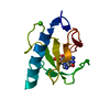
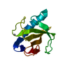
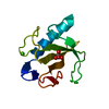
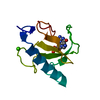
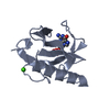
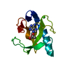
| Title | RIBONUCLEASE T1 WITH FREE RECOGNITION AND CATALYTIC SITE: CRYSTAL STRUCTURE ANALYSIS AT 1.5 ANGSTROMS RESOLUTION | ||||||
 Components Components | RIBONUCLEASE T1 | ||||||
 Keywords Keywords | HYDROLASE(ENDORIBONUCLEASE) | ||||||
| Function / homology |  Function and homology information Function and homology informationhyphal tip / ribonuclease T1 activity / ribonuclease T1 / cell septum / endonuclease activity / lyase activity / RNA bindingSimilarity search - Function | ||||||
| Biological species |  Aspergillus oryzae (mold) Aspergillus oryzae (mold) | ||||||
| Method | X-RAY DIFFRACTION / Resolution: 1.5 Å | ||||||
 Authors Authors | Martinez-Oyanedel, J. / Heinemann, U. / Saenger, W. | ||||||
 Citation Citation | Journal: J.Mol.Biol. / Year: 1991 Title: Ribonuclease T1 with free recognition and catalytic site: crystal structure analysis at 1.5 A resolution. Authors: Martinez-Oyanedel, J. / Choe, H.W. / Heinemann, U. / Saenger, W. #1: Journal: Biochemistry / Year: 1989Title: Crystal Structure of Guanosine-Free Ribonuclease T1, Complexed with Vanadate(V), Suggests Conformational Change Upon Substrate Binding Authors: Kostrewa, D. / Choe, H.-W. / Heinemann, U. / Saenger, W. #2: Journal: J.Biol.Chem. / Year: 1988Title: Three-Dimensional Structure of the Ribonuclease T1(Asterisk)2'-Gmp Complex at 1.9-Angstroms Resolution Authors: Arni, R. / Heinemann, U. / Tokuoka, R. / Saenger, W. #3: Journal: Nature / Year: 1982Title: Specific Protein-Nucleic Acid Recognition in Ribonuclease T1-2'-Guanylic Acid Complex. An X-Ray Study Authors: Heinemann, U. / Saenger, W. | ||||||
| History |
|
- Structure visualization
| Structure viewer | Molecule: MolmilJmol/JSmol |
|---|
- Downloads & links
-Download
| PDBx/mmCIF format | 9rnt.cif.gz | 31.5 KB | Display | PDBx/mmCIF format |
|---|---|---|---|---|
| PDB format | pdb9rnt.ent.gz | 23.6 KB | Display | PDB format |
| PDBx/mmJSON format | 9rnt.json.gz | Tree view | PDBx/mmJSON format | |
| Others |  Other downloads Other downloads |
-Validation report
| Arichive directory | https://data.pdbj.org/pub/pdb/validation_reports/rn/9rntftp://data.pdbj.org/pub/pdb/validation_reports/rn/9rnt | HTTPS FTP |
|---|
-Related structure data
| Similar structure data |
|---|


-Links
 PDBj
PDBj- Assembly
| Deposited unit | 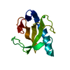
| ||||||||
|---|---|---|---|---|---|---|---|---|---|
| 1 |
| ||||||||
| Unit cell |
| ||||||||
| Atom site foot note | 1: RESIDUES PRO 39 AND PRO 55 ARE CIS PROLINES. |
-Components
| #1: Protein | Mass: 11094.694 Da / Num. of mol.: 1 Source method: isolated from a genetically manipulated source Source: (gene. exp.) Aspergillus oryzae (mold) / References: UniProt: P00651, EC: 3.1.27.3 |
|---|---|
| #2: Chemical | ChemComp-CA /   Mass: 40.078 Da / Num. of mol.: 1 / Source method: obtained synthetically / Formula: Ca Mass: 40.078 Da / Num. of mol.: 1 / Source method: obtained synthetically / Formula: Ca |
| #3: Water | ChemComp-HOH / Water Mass: 18.015 Da / Num. of mol.: 121 / Source method: isolated from a natural source / Formula: H2O Mass: 18.015 Da / Num. of mol.: 121 / Source method: isolated from a natural source / Formula: H2O |
-Experimental details
-Experiment
| Experiment | Method: X-RAY DIFFRACTION |
|---|
- Sample preparation
| Crystal | Density Matthews: 2.09 Å3/Da / Density % sol: 41.23 % | ||||||||||||||||||||||||||||||
|---|---|---|---|---|---|---|---|---|---|---|---|---|---|---|---|---|---|---|---|---|---|---|---|---|---|---|---|---|---|---|---|
| Crystal grow | *PLUS pH: 7 / Method: vapor diffusion | ||||||||||||||||||||||||||||||
| Components of the solutions | *PLUS
|
-Data collection
| Reflection | *PLUS Highest resolution: 1.5 Å / Lowest resolution: 10 Å / Num. obs: 13115 / % possible obs: 84.5 % / Num. measured all: 17624 / Rmerge F obs: 1 |
|---|
- Processing
| Software | Name: PROFFT / Classification: refinement | ||||||||||||||||||||||||||||||||||||||||||||||||||||||||||||||||||||||||||||||||||||
|---|---|---|---|---|---|---|---|---|---|---|---|---|---|---|---|---|---|---|---|---|---|---|---|---|---|---|---|---|---|---|---|---|---|---|---|---|---|---|---|---|---|---|---|---|---|---|---|---|---|---|---|---|---|---|---|---|---|---|---|---|---|---|---|---|---|---|---|---|---|---|---|---|---|---|---|---|---|---|---|---|---|---|---|---|---|
| Refinement | Resolution: 1.5→6 Å / σ(F): 0 /
| ||||||||||||||||||||||||||||||||||||||||||||||||||||||||||||||||||||||||||||||||||||
| Refinement step | Cycle: LAST / Resolution: 1.5→6 Å
| ||||||||||||||||||||||||||||||||||||||||||||||||||||||||||||||||||||||||||||||||||||
| Refine LS restraints |
|
