National Natural Science Foundation of China (NSFC)
China
Citation
Journal: J Virol / Year: 2024 Title: African swine fever virus A137R assembles into a dodecahedron cage. Authors: Changyao Li / Mingzhu Jia / Tianjiao Hao / Qi Peng / Ruchao Peng / Yan Chai / Yi Shi / Hao Song / George F Gao / Abstract: African swine fever (ASF) is a highly contagious viral disease that affects domestic and wild pigs. The causative agent of ASF is African swine fever virus (ASFV), a large double-stranded DNA virus ...African swine fever (ASF) is a highly contagious viral disease that affects domestic and wild pigs. The causative agent of ASF is African swine fever virus (ASFV), a large double-stranded DNA virus with a complex virion structure. Among the various proteins encoded by ASFV, A137R is a crucial structural protein associated with its virulence. However, the structure and molecular mechanisms underlying the functions of A137R remain largely unknown. In this study, we present the structure of A137R determined by cryogenic electron microscopy single-particle reconstruction, which reveals that A137R self-oligomerizes to form a dodecahedron-shaped cage composed of 60 polymers. The dodecahedron is literally equivalent to a 1 icosahedron where the icosahedral vertexes are located in the center of each dodecahedral facet. Within each facet, five A137R protomers are arranged in a head-to-tail orientation with a long N-terminal helix forming the edge through which adjacent facets stitch together to form the dodecahedral cage. Combining structural analysis and biochemical evidence, we demonstrate that the N-terminal domain of A137R is crucial and sufficient for mediating the assembly of the dodecahedron. These findings imply the role of A137R cage as a core component in the icosahedral ASFV virion and suggest a promising molecular scaffold for nanotechnology applications. IMPORTANCE: African swine fever (ASF) is a lethal viral disease of pigs caused by African swine fever virus (ASFV). No commercial vaccines and antiviral treatments are available for the prevention ...IMPORTANCE: African swine fever (ASF) is a lethal viral disease of pigs caused by African swine fever virus (ASFV). No commercial vaccines and antiviral treatments are available for the prevention and control of the disease. A137R is a structural protein of ASFV that is associated with its virulence. The discovery of the dodecahedron-shaped cage structure of A137R in this study is of great importance in understanding ASFV pathogenicity. This finding sheds light on the molecular mechanisms underlying the functions of A137R. Furthermore, the dodecahedral cage formed by A137R shows promise as a molecular scaffold for nanoparticle vectors. Overall, this study provides valuable insights into the structure and function of A137R, contributing to our understanding of ASFV and potentially opening up new avenues for the development of vaccines or treatments for ASF.
In the structure databanks used in Yorodumi, some data are registered as the other names, "COVID-19 virus" and "2019-nCoV". Here are the details of the virus and the list of structure data.
Jan 31, 2019. EMDB accession codes are about to change! (news from PDBe EMDB page)
EMDB accession codes are about to change! (news from PDBe EMDB page)
The allocation of 4 digits for EMDB accession codes will soon come to an end. Whilst these codes will remain in use, new EMDB accession codes will include an additional digit and will expand incrementally as the available range of codes is exhausted. The current 4-digit format prefixed with “EMD-” (i.e. EMD-XXXX) will advance to a 5-digit format (i.e. EMD-XXXXX), and so on. It is currently estimated that the 4-digit codes will be depleted around Spring 2019, at which point the 5-digit format will come into force.
The EM Navigator/Yorodumi systems omit the EMD- prefix.
Related info.:Q: What is EMD? / ID/Accession-code notation in Yorodumi/EM Navigator
Yorodumi is a browser for structure data from EMDB, PDB, SASBDB, etc.
This page is also the successor to EM Navigator detail page, and also detail information page/front-end page for Omokage search.
The word "yorodu" (or yorozu) is an old Japanese word meaning "ten thousand". "mi" (miru) is to see.
Related info.:EMDB / PDB / SASBDB / Comparison of 3 databanks / Yorodumi Search / Aug 31, 2016. New EM Navigator & Yorodumi / Yorodumi Papers / Jmol/JSmol / Function and homology information / Changes in new EM Navigator and Yorodumi
 Movie
Movie Controller


 Open data
 Basic information
 Components
 Keywords
 VIRAL PROTEIN /
  Function and homology information

 Authors
 China, 1items
  Citation
 Structure visualization
 Downloads & links
 Other downloads
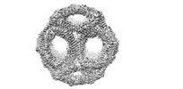
 PDBj
PDBj Assembly

 Sample preparation
 Electron microscopy imaging
 Processing