 Movie
Movie Controller


+ Open data

- Basic information
| Entry | Database: PDB / ID: 8x90 | ||||||
|---|---|---|---|---|---|---|---|
| Title | P/Q type calcium channel | ||||||
 Components Components | (Voltage-dependent ...) x 3 | ||||||
 Keywords Keywords |  MEMBRANE PROTEIN / voltage-gated calcium channel MEMBRANE PROTEIN / voltage-gated calcium channel | ||||||
| Function / homology |  Function and homology information Function and homology informationregulation of membrane repolarization during action potential / Presynaptic depolarization and calcium channel opening / positive regulation of high voltage-gated calcium channel activity / calcium ion transmembrane transport via high voltage-gated calcium channel / membrane depolarization during bundle of His cell action potential / L-type voltage-gated calcium channel complex / cardiac muscle cell action potential involved in contraction / high voltage-gated calcium channel activity / NCAM1 interactions / regulation of ventricular cardiac muscle cell membrane repolarization ...regulation of membrane repolarization during action potential / Presynaptic depolarization and calcium channel opening / positive regulation of high voltage-gated calcium channel activity / calcium ion transmembrane transport via high voltage-gated calcium channel / membrane depolarization during bundle of His cell action potential / L-type voltage-gated calcium channel complex / cardiac muscle cell action potential involved in contraction / high voltage-gated calcium channel activity / NCAM1 interactions / regulation of ventricular cardiac muscle cell membrane repolarization / calcium ion transport into cytosol / regulation of calcium ion transmembrane transport via high voltage-gated calcium channel / syntaxin binding / neuromuscular junction development / voltage-gated calcium channel complex / neuronal dense core vesicle / regulation of heart rate by cardiac conduction / calcium ion import across plasma membrane / calcium channel regulator activity / regulation of calcium ion transport / response to amyloid-beta / voltage-gated calcium channel activity / sarcoplasmic reticulum / cell projection / Regulation of insulin secretion / protein localization to plasma membrane / calcium ion transmembrane transport / modulation of chemical synaptic transmission / Adrenaline,noradrenaline inhibits insulin secretion / cellular response to amyloid-beta / calcium ion transport / amyloid-beta binding / positive regulation of cytosolic calcium ion concentration / T cell receptor signaling pathway / chemical synaptic transmission / neuronal cell body / synapse / extracellular exosome / membrane / metal ion binding / nucleus / plasma membrane / cytosol / cytoplasmSimilarity search - Function | ||||||
| Biological species |  Homo sapiens (human) Homo sapiens (human) | ||||||
| Method | ELECTRON MICROSCOPY / single particle reconstruction / cryo EM / Resolution: 2.95 Å | ||||||
 Authors Authors | Yan, N. / Li, Z. / Cong, Y. / Wu, T. / Wang, T. | ||||||
| Funding support |  China, 1items China, 1items
| ||||||
 Citation Citation | Journal: Cell Res / Year: 2024 Title: Structural basis for different ω-agatoxin IVA sensitivities of the P-type and Q-type Ca2.1 channels. Authors: Zhangqiang Li / Ye Cong / Tong Wu / Tongtong Wang / Xinyao Lou / Xinyu Yang / Nieng Yan / | ||||||
| History |
|
- Structure visualization
| Structure viewer | Molecule: MolmilJmol/JSmol |
|---|
- Downloads & links
-Download
| PDBx/mmCIF format | 8x90.cif.gz | 536.6 KB | Display | PDBx/mmCIF format |
|---|---|---|---|---|
| PDB format | pdb8x90.ent.gz | 412 KB | Display | PDB format |
| PDBx/mmJSON format | 8x90.json.gz | Tree view | PDBx/mmJSON format | |
| Others |  Other downloads Other downloads |
-Validation report
| Arichive directory | https://data.pdbj.org/pub/pdb/validation_reports/x9/8x90ftp://data.pdbj.org/pub/pdb/validation_reports/x9/8x90 | HTTPS FTP |
|---|
-Related structure data
| Related structure data |  38158MC 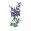 8x91C 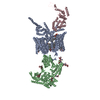 8x93C M: map data used to model this data C: citing same article ( |
|---|---|
| Similar structure data |


-Links
 PDBj
PDBj


- Assembly
| Deposited unit | 
|
|---|---|
| 1 |
|
-Components
-Voltage-dependent ... , 3 types, 3 molecules ABC
| #1: Protein | Mass: 287534.312 Da / Num. of mol.: 1 Source method: isolated from a genetically manipulated source Source: (gene. exp.) Homo sapiens (human) / Gene: CACNA1A, CACH4, CACN3, CACNL1A4 / Production host: Homo sapiens (human) / References: UniProt: O00555 |
|---|---|
| #2: Protein | Voltage-gated calcium channel / Voltage-gated calcium channel subunit alpha-2/delta-1 Mass: 126316.148 Da / Num. of mol.: 1 Source method: isolated from a genetically manipulated source Source: (gene. exp.) Homo sapiens (human) / Gene: CACNA2D1, CACNL2A, CCHL2A, MHS3 / Production host: Homo sapiens (human) / References: UniProt: P54289 |
| #3: Protein | Mass: 56231.570 Da / Num. of mol.: 1 Source method: isolated from a genetically manipulated source Source: (gene. exp.) Homo sapiens (human) / Gene: CACNB3, CACNLB3 / Production host: Homo sapiens (human) / References: UniProt: P54284 |
-Sugars , 4 types, 7 molecules 
| #4: Polysaccharide | 2-acetamido-2-deoxy-beta-D-glucopyranose-(1-4)-2-acetamido-2-deoxy-beta-D-glucopyranose-(1-4)-2- ...2-acetamido-2-deoxy-beta-D-glucopyranose-(1-4)-2-acetamido-2-deoxy-beta-D-glucopyranose-(1-4)-2-acetamido-2-deoxy-beta-D-glucopyranose / Mass: 627.594 Da / Num. of mol.: 1 Source method: isolated from a genetically manipulated source | ||||
|---|---|---|---|---|---|
| #5: Polysaccharide | / Mass: 424.401 Da / Num. of mol.: 3 Source method: isolated from a genetically manipulated source #6: Polysaccharide | 2-acetamido-2-deoxy-beta-D-glucopyranose-(1-4)-2-acetamido-2-deoxy-beta-D-glucopyranose-(1-4)-2- ...2-acetamido-2-deoxy-beta-D-glucopyranose-(1-4)-2-acetamido-2-deoxy-beta-D-glucopyranose-(1-4)-2-acetamido-2-deoxy-beta-D-glucopyranose-(1-4)-2-acetamido-2-deoxy-beta-D-glucopyranose | / Mass: 830.786 Da / Num. of mol.: 1Source method: isolated from a genetically manipulated source #11: Sugar | N-Acetylglucosamine Type: D-saccharide, beta linking / Mass: 221.208 Da / Num. of mol.: 2 / Source method: obtained synthetically / Formula: C8H15NO6 Type: D-saccharide, beta linking / Mass: 221.208 Da / Num. of mol.: 2 / Source method: obtained synthetically / Formula: C8H15NO6 |
-Non-polymers , 6 types, 14 molecules 


| #7: Chemical | ChemComp-PS1 /  Mass: 566.642 Da / Num. of mol.: 1 / Source method: obtained synthetically / Formula: C26H49NO10P Mass: 566.642 Da / Num. of mol.: 1 / Source method: obtained synthetically / Formula: C26H49NO10P | ||||||||
|---|---|---|---|---|---|---|---|---|---|
| #8: Chemical | ChemComp-CLR / Cholesterol Mass: 386.654 Da / Num. of mol.: 5 / Source method: obtained synthetically / Formula: C27H46O Mass: 386.654 Da / Num. of mol.: 5 / Source method: obtained synthetically / Formula: C27H46O#9: Chemical | ChemComp-PT5 / [( | Phosphatidylinositol 4,5-bisphosphate Mass: 1047.088 Da / Num. of mol.: 1 / Source method: obtained synthetically / Formula: C47H85O19P3 / Comment: phospholipid*YM Mass: 1047.088 Da / Num. of mol.: 1 / Source method: obtained synthetically / Formula: C47H85O19P3 / Comment: phospholipid*YM#10: Chemical |  Mass: 40.078 Da / Num. of mol.: 2 / Source method: obtained synthetically / Formula: Ca Mass: 40.078 Da / Num. of mol.: 2 / Source method: obtained synthetically / Formula: Ca#12: Chemical |  Mass: 486.726 Da / Num. of mol.: 3 / Source method: obtained synthetically / Formula: C31H50O4 Mass: 486.726 Da / Num. of mol.: 3 / Source method: obtained synthetically / Formula: C31H50O4#13: Chemical | Phosphatidylethanolamine Mass: 748.065 Da / Num. of mol.: 2 / Source method: obtained synthetically / Formula: C41H82NO8P / Comment: phospholipid*YM Mass: 748.065 Da / Num. of mol.: 2 / Source method: obtained synthetically / Formula: C41H82NO8P / Comment: phospholipid*YM |
-Details
| Has ligand of interest | N |
|---|
-Experimental details
-Experiment
| Experiment | Method: ELECTRON MICROSCOPY |
|---|---|
| EM experiment | Aggregation state: PARTICLE / 3D reconstruction method: single particle reconstruction |
- Sample preparation
| Component | Name: Voltage-gated P/Q type calcium channel / Type: COMPLEX / Entity ID: #1-#3 / Source: RECOMBINANT |
|---|---|
| Source (natural) | Organism: Homo sapiens (human) |
| Source (recombinant) | Organism: Homo sapiens (human) |
| Buffer solution | pH: 8 |
| Specimen | Embedding applied: NO / Shadowing applied: NO / Staining applied: NO / Vitrification applied: YES |
| Vitrification | Cryogen name: ETHANE |
- Electron microscopy imaging
| Experimental equipment |  Model: Titan Krios / Image courtesy: FEI Company |
|---|---|
| Microscopy | Model: FEI TITAN KRIOS |
| Electron gun | Electron source: FIELD EMISSION GUN / Accelerating voltage: 300 kV / Illumination mode: SPOT SCAN |
| Electron lens | Mode: BRIGHT FIELDBright-field microscopy / Nominal defocus max: 1800 nm / Nominal defocus min: 1300 nm |
| Image recording | Electron dose: 50 e/Å2 / Film or detector model: GATAN K3 (6k x 4k) |
- Processing
| EM software | Name: PHENIX / Version: 1.19.2_4158: / Category: model refinement | ||||||||||||||||||||||||
|---|---|---|---|---|---|---|---|---|---|---|---|---|---|---|---|---|---|---|---|---|---|---|---|---|---|
| CTF correction | Type: PHASE FLIPPING AND AMPLITUDE CORRECTION | ||||||||||||||||||||||||
| 3D reconstruction | Resolution: 2.95 Å / Resolution method: FSC 0.143 CUT-OFF / Num. of particles: 118997 / Symmetry type: POINT | ||||||||||||||||||||||||
| Refine LS restraints |
|
