 Movie
Movie Controller


+ Open data

- Basic information
| Entry | Database: PDB / ID: 8vgd | ||||||
|---|---|---|---|---|---|---|---|
| Title | Complex of ExbD with D-box peptide: Tetragonal form | ||||||
 Components Components |
| ||||||
 Keywords Keywords |  TRANSPORT PROTEIN / TonB / TonB-dependent transport / bacterial motor TRANSPORT PROTEIN / TonB / TonB-dependent transport / bacterial motor | ||||||
| Function / homology |  Function and homology information Function and homology informationreceptor-mediated bacteriophage irreversible attachment to host cell / colicin transport / energy transducer activity / cell envelope / cobalamin transport / siderophore transport / intracellular monoatomic cation homeostasis / plasma membrane protein complex / transmembrane transporter complex / transmembrane transporter activity ...receptor-mediated bacteriophage irreversible attachment to host cell / colicin transport / energy transducer activity / cell envelope / cobalamin transport / siderophore transport / intracellular monoatomic cation homeostasis / plasma membrane protein complex / transmembrane transporter complex / transmembrane transporter activity / cell outer membrane / transmembrane transport / protein transport / outer membrane-bounded periplasmic space / intracellular iron ion homeostasis / protein domain specific binding / membrane / plasma membraneSimilarity search - Function | ||||||
| Biological species |  Escherichia coli (E. coli) Escherichia coli (E. coli) | ||||||
| Method | X-RAY DIFFRACTION / SYNCHROTRON / MOLECULAR REPLACEMENT / Resolution: 1.42 Å | ||||||
 Authors Authors | Loll, P.J. | ||||||
| Funding support | 1items
| ||||||
 Citation Citation | Journal: J.Biol.Chem. / Year: 2024 Title: Discovery and structural characterization of the D-box, a conserved TonB motif that couples an inner-membrane motor to outer-membrane transport. Authors: Loll, P.J. / Grasty, K.C. / Shultis, D.D. / Guzman, N.J. / Wiener, M.C. | ||||||
| History |
|
- Structure visualization
| Structure viewer | Molecule: MolmilJmol/JSmol |
|---|
- Downloads & links
-Download
| PDBx/mmCIF format | 8vgd.cif.gz | 102.8 KB | Display | PDBx/mmCIF format |
|---|---|---|---|---|
| PDB format | pdb8vgd.ent.gz | 80 KB | Display | PDB format |
| PDBx/mmJSON format | 8vgd.json.gz | Tree view | PDBx/mmJSON format | |
| Others |  Other downloads Other downloads |
-Validation report
| Arichive directory | https://data.pdbj.org/pub/pdb/validation_reports/vg/8vgdftp://data.pdbj.org/pub/pdb/validation_reports/vg/8vgd | HTTPS FTP |
|---|
-Related structure data
| Related structure data | 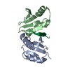 8vgcC C: citing same article ( |
|---|---|
| Similar structure data | |
| Experimental dataset #1 | Data reference: 10.5281/zenodo.10574068 / Data set type: diffraction image data |
-Links
 PDBj
PDBj- Assembly
| Deposited unit | 
| ||||||||
|---|---|---|---|---|---|---|---|---|---|
| 1 |
| ||||||||
| Unit cell |
|
-Components
| #1: Protein | Mass: 9238.591 Da / Num. of mol.: 2 / Fragment: periplasmic domain Source method: isolated from a genetically manipulated source Source: (gene. exp.) Escherichia coli (E. coli) / Gene: exbD, EIMP300_23080 / Plasmid: pETHSUL / Production host: Escherichia coli BL21(DE3) (bacteria) / References: UniProt: A0A8S0FLD5#2: Protein/peptide | | Mass: 975.159 Da / Num. of mol.: 1 / Fragment: D-box peptide / Source method: obtained synthetically / Details: D-box peptide from TonB / Source: (synth.) Escherichia coli (E. coli) / References: UniProt: P02929#3: Water | ChemComp-HOH / | Water Mass: 18.015 Da / Num. of mol.: 50 / Source method: isolated from a natural source / Formula: H2O Mass: 18.015 Da / Num. of mol.: 50 / Source method: isolated from a natural source / Formula: H2O |
|---|
-Experimental details
-Experiment
| Experiment | Method: X-RAY DIFFRACTION / Number of used crystals: 1 |
|---|
- Sample preparation
| Crystal | Density Matthews: 1.93 Å3/Da / Density % sol: 36.39 % |
|---|---|
| Crystal grow | Temperature: 277 K / Method: microbatch / pH: 5.8 Details: 2M ammonium sulfate in 0.1 M sodium citrate, pH 5.8; microbatch under Al's oil |
-Data collection
| Diffraction | Mean temperature: 100 K / Serial crystal experiment: N | ||||||||||||||||||||||||||||||||||||||||||||||||||||||||||||||||||||||||||||||||||||||||||||||||||||||||||||||||||||||||||||||
|---|---|---|---|---|---|---|---|---|---|---|---|---|---|---|---|---|---|---|---|---|---|---|---|---|---|---|---|---|---|---|---|---|---|---|---|---|---|---|---|---|---|---|---|---|---|---|---|---|---|---|---|---|---|---|---|---|---|---|---|---|---|---|---|---|---|---|---|---|---|---|---|---|---|---|---|---|---|---|---|---|---|---|---|---|---|---|---|---|---|---|---|---|---|---|---|---|---|---|---|---|---|---|---|---|---|---|---|---|---|---|---|---|---|---|---|---|---|---|---|---|---|---|---|---|---|---|---|
| Diffraction source | Source: SYNCHROTRON / Site: NSLS-II  / Beamline: 17-ID-1 / Wavelength: 0.92011 Å / Beamline: 17-ID-1 / Wavelength: 0.92011 Å | ||||||||||||||||||||||||||||||||||||||||||||||||||||||||||||||||||||||||||||||||||||||||||||||||||||||||||||||||||||||||||||||
| Detector | Type: DECTRIS EIGER X 9M / Detector: PIXEL / Date: Oct 14, 2021 | ||||||||||||||||||||||||||||||||||||||||||||||||||||||||||||||||||||||||||||||||||||||||||||||||||||||||||||||||||||||||||||||
| Radiation | Protocol: SINGLE WAVELENGTH / Monochromatic (M) / Laue (L): M / Scattering type: x-ray | ||||||||||||||||||||||||||||||||||||||||||||||||||||||||||||||||||||||||||||||||||||||||||||||||||||||||||||||||||||||||||||||
| Radiation wavelength | Wavelength: 0.92011 Å / Relative weight: 1 | ||||||||||||||||||||||||||||||||||||||||||||||||||||||||||||||||||||||||||||||||||||||||||||||||||||||||||||||||||||||||||||||
| Reflection | Resolution: 1.42→26.4 Å / Num. obs: 27989 / % possible obs: 100 % / Redundancy: 14.4 % / CC1/2: 0.998 / Rmerge(I) obs: 0.08 / Rrim(I) all: 0.083 / Net I/σ(I): 13.6 | ||||||||||||||||||||||||||||||||||||||||||||||||||||||||||||||||||||||||||||||||||||||||||||||||||||||||||||||||||||||||||||||
| Reflection shell |
|
- Processing
| Software |
| ||||||||||||||||
|---|---|---|---|---|---|---|---|---|---|---|---|---|---|---|---|---|---|
| Refinement | Method to determine structure: MOLECULAR REPLACEMENT / Resolution: 1.42→24.63 Å / Cross valid method: FREE R-VALUE
| ||||||||||||||||
| Refinement step | Cycle: LAST / Resolution: 1.42→24.63 Å
|
