 Movie
Movie Controller


+ Open data

- Basic information
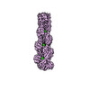
| Entry | Database: PDB / ID: 8v2o | ||||||
|---|---|---|---|---|---|---|---|
| Title | Cryo-EM Structure of Wildtype Smooth Muscle Gamma Actin (ACTG2) | ||||||
 Components Components | Actin, gamma-enteric smooth muscle | ||||||
 Keywords Keywords | STRUCTURAL PROTEIN / Filament / Actin / Smooth Muscle / CYTOSOLIC PROTEIN | ||||||
| Function / homology |  Function and homology information Function and homology informationmyosin filament / mesenchyme migration / Smooth Muscle Contraction / filopodium / cell periphery / Hydrolases; Acting on acid anhydrides; Acting on acid anhydrides to facilitate cellular and subcellular movement / lamellipodium / cell body / blood microparticle / cytoskeleton ...myosin filament / mesenchyme migration / Smooth Muscle Contraction / filopodium / cell periphery / Hydrolases; Acting on acid anhydrides; Acting on acid anhydrides to facilitate cellular and subcellular movement / lamellipodium / cell body / blood microparticle / cytoskeleton / hydrolase activity / positive regulation of gene expression / extracellular space / extracellular exosome / ATP binding / cytosol / cytoplasmSimilarity search - Function | ||||||
| Biological species |  Homo sapiens (human) Homo sapiens (human) | ||||||
| Method | ELECTRON MICROSCOPY / helical reconstruction / cryo EM / Resolution: 2.45 Å | ||||||
 Authors Authors | Palmer, N.J. / Carman, P.J. / Ceron, R.H. / Dominguez, R. | ||||||
| Funding support |  United States, 1items United States, 1items
| ||||||
 Citation Citation | Journal: To Be Published Title: Molecular Mechanisms of ACTG2 Mutations Linked to Visceral Myopathy Authors: Ceron, R.H. / Baez-Cruz, F.A. / Palmer, N.J. / Carman, P.J. / Boczkowska, M. / Heuckeroth, R.O. / Ostap, E.M. / Dominguez, R. | ||||||
| History |
|
- Structure visualization
| Structure viewer | Molecule: MolmilJmol/JSmol |
|---|
- Downloads & links
-Download
| PDBx/mmCIF format | 8v2o.cif.gz | 362.5 KB | Display | PDBx/mmCIF format |
|---|---|---|---|---|
| PDB format | pdb8v2o.ent.gz | 295.8 KB | Display | PDB format |
| PDBx/mmJSON format | 8v2o.json.gz | Tree view | PDBx/mmJSON format | |
| Others |  Other downloads Other downloads |
-Validation report
| Arichive directory | https://data.pdbj.org/pub/pdb/validation_reports/v2/8v2oftp://data.pdbj.org/pub/pdb/validation_reports/v2/8v2o | HTTPS FTP |
|---|
-Related structure data
| Related structure data |  42918MC 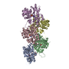 8v30C M: map data used to model this data C: citing same article ( |
|---|---|
| Similar structure data |
-Links
 PDBj
PDBj


- Assembly
| Deposited unit | 
|
|---|---|
| 1 |
|
-Components
| #1: Protein | / Alpha-actin-3 / Gamma-2-actin / Smooth muscle gamma-actin / ACTG2 Mass: 41935.816 Da / Num. of mol.: 5 Source method: isolated from a genetically manipulated source Source: (gene. exp.) Homo sapiens (human) / Tissue: Smooth Muscle / Gene: ACTG2, ACTA3, ACTL3, ACTSG / Cell line (production host): Expi293F / Production host: Homo sapiens (human)References: UniProt: P63267, Hydrolases; Acting on acid anhydrides; Acting on acid anhydrides to facilitate cellular and subcellular movement#2: Chemical | ChemComp-ADP / Adenosine diphosphate  Mass: 427.201 Da / Num. of mol.: 5 / Source method: obtained synthetically / Formula: C10H15N5O10P2 / Comment: ADP, energy-carrying molecule*YM Mass: 427.201 Da / Num. of mol.: 5 / Source method: obtained synthetically / Formula: C10H15N5O10P2 / Comment: ADP, energy-carrying molecule*YM#3: Chemical | ChemComp-MG /   Mass: 24.305 Da / Num. of mol.: 5 / Source method: obtained synthetically / Formula: Mg Mass: 24.305 Da / Num. of mol.: 5 / Source method: obtained synthetically / Formula: MgHas ligand of interest | N | Sequence details | Numbering of the actin chains is shifted -1 from the medical ACTG2 literature to match the ...Numbering of the actin chains is shifted -1 from the medical ACTG2 literature to match the conventional numbering | |
|---|
-Experimental details
-Experiment
| Experiment | Method: ELECTRON MICROSCOPY |
|---|---|
| EM experiment | Aggregation state: FILAMENT / 3D reconstruction method: helical reconstruction |
- Sample preparation
| Component | Name: ACTG2 filament / Type: COMPLEX / Details: ACTG2 filaments, purified from Expi293 cells / Entity ID: #1 / Source: RECOMBINANT | |||||||||||||||||||||||||||||||||||
|---|---|---|---|---|---|---|---|---|---|---|---|---|---|---|---|---|---|---|---|---|---|---|---|---|---|---|---|---|---|---|---|---|---|---|---|---|
| Molecular weight | Units: KILODALTONS/NANOMETER / Experimental value: NO | |||||||||||||||||||||||||||||||||||
| Source (natural) | Organism: Homo sapiens (human) / Tissue: Smooth | |||||||||||||||||||||||||||||||||||
| Source (recombinant) | Organism: Homo sapiens (human) / Cell: Expi293F | |||||||||||||||||||||||||||||||||||
| Buffer solution | pH: 8 / Details: Actin F-buffer | |||||||||||||||||||||||||||||||||||
| Buffer component |
| |||||||||||||||||||||||||||||||||||
| Specimen | Conc.: 0.168 mg/ml / Embedding applied: NO / Shadowing applied: NO / Staining applied: NO / Vitrification applied: YES / Details: ACTG2 F-Actin in the ADP state. | |||||||||||||||||||||||||||||||||||
| Specimen support | Grid material: COPPER / Grid mesh size: 200 divisions/in. / Grid type: Quantifoil R1.2/1.3 | |||||||||||||||||||||||||||||||||||
| Vitrification | Instrument: FEI VITROBOT MARK III / Cryogen name: ETHANE / Humidity: 100 % / Chamber temperature: 277 K / Details: Blot Force: 0 Blot Time: 2.5 s |
- Electron microscopy imaging
| Experimental equipment |  Model: Titan Krios / Image courtesy: FEI Company |
|---|---|
| Microscopy | Model: FEI TITAN KRIOS |
| Electron gun | Electron source: FIELD EMISSION GUN / Accelerating voltage: 300 kV / Illumination mode: SPOT SCAN |
| Electron lens | Mode: BRIGHT FIELDBright-field microscopy / Nominal magnification: 81000 X / Nominal defocus max: 2500 nm / Nominal defocus min: 500 nm / C2 aperture diameter: 100 µm |
| Specimen holder | Cryogen: NITROGEN / Specimen holder model: FEI TITAN KRIOS AUTOGRID HOLDER |
| Image recording | Electron dose: 45 e/Å2 / Film or detector model: GATAN K3 (6k x 4k) / Num. of real images: 526 |
- Processing
| EM software |
| |||||||||||||||||||||||||||||||||||
|---|---|---|---|---|---|---|---|---|---|---|---|---|---|---|---|---|---|---|---|---|---|---|---|---|---|---|---|---|---|---|---|---|---|---|---|---|
| CTF correction | Type: PHASE FLIPPING AND AMPLITUDE CORRECTION | |||||||||||||||||||||||||||||||||||
| Helical symmerty | Angular rotation/subunit: -166.6 ° / Axial rise/subunit: 27.5 Å / Axial symmetry: C1 | |||||||||||||||||||||||||||||||||||
| 3D reconstruction | Resolution: 2.45 Å / Resolution method: FSC 0.143 CUT-OFF / Num. of particles: 473627 / Symmetry type: HELICAL | |||||||||||||||||||||||||||||||||||
| Atomic model building | Protocol: RIGID BODY FIT / Space: REAL | |||||||||||||||||||||||||||||||||||
| Refine LS restraints |
|
