+ Open data
- Basic information
| Entry | Database: PDB / ID: 8uvt | ||||||
|---|---|---|---|---|---|---|---|
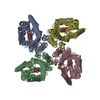
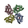
| Title | Structure of the insect gustatory receptor Gr9 from Bombyx mori | ||||||
 Components Components | Gustatory receptor | ||||||
 Keywords Keywords |  TRANSPORT PROTEIN / Gustatory receptor / TRANSPORT PROTEIN / Gustatory receptor /  Ion channel / Sugar-binding / Ion channel / Sugar-binding /  Fructose Fructose | ||||||
| Function / homology | 7TM chemoreceptor / 7tm Chemosensory receptor / sensory perception of taste /  signal transduction / signal transduction /  plasma membrane / Gustatory receptor plasma membrane / Gustatory receptor Function and homology information Function and homology information | ||||||
| Biological species |   Bombyx mori (domestic silkworm) Bombyx mori (domestic silkworm) | ||||||
| Method |  ELECTRON MICROSCOPY / ELECTRON MICROSCOPY /  single particle reconstruction / single particle reconstruction /  cryo EM / Resolution: 2.9 Å cryo EM / Resolution: 2.9 Å | ||||||
 Authors Authors | Gomes, J.V. / Butterwick, J.A. | ||||||
| Funding support | 1items
| ||||||
 Citation Citation |  Journal: Nature / Year: 2024 Journal: Nature / Year: 2024Title: The molecular basis of sugar detection by an insect taste receptor. Authors: João Victor Gomes / Shivinder Singh-Bhagania / Matthew Cenci / Carlos Chacon Cordon / Manjodh Singh / Joel A Butterwick /  Abstract: Animals crave sugars because of their energy potential and the pleasurable sensation of tasting sweetness. Yet all sugars are not metabolically equivalent, requiring mechanisms to detect and ...Animals crave sugars because of their energy potential and the pleasurable sensation of tasting sweetness. Yet all sugars are not metabolically equivalent, requiring mechanisms to detect and differentiate between chemically similar sweet substances. Insects use a family of ionotropic gustatory receptors to discriminate sugars, each of which is selectively activated by specific sweet molecules. Here, to gain insight into the molecular basis of sugar selectivity, we determined structures of Gr9, a gustatory receptor from the silkworm Bombyx mori (BmGr9), in the absence and presence of its sole activating ligand, D-fructose. These structures, along with structure-guided mutagenesis and functional assays, illustrate how D-fructose is enveloped by a ligand-binding pocket that precisely matches the overall shape and pattern of chemical groups in D-fructose. However, our computational docking and experimental binding assays revealed that other sugars also bind BmGr9, yet they are unable to activate the receptor. We determined the structure of BmGr9 in complex with one such non-activating sugar, L-sorbose. Although both sugars bind a similar position, only D-fructose is capable of engaging a bridge of two conserved aromatic residues that connects the pocket to the pore helix, inducing a conformational change that allows the ion-conducting pore to open. Thus, chemical specificity does not depend solely on the selectivity of the ligand-binding pocket, but it is an emergent property arising from a combination of receptor-ligand interactions and allosteric coupling. Our results support a model whereby coarse receptor tuning is derived from the size and chemical characteristics of the pocket, whereas fine-tuning of receptor activation is achieved through the selective engagement of an allosteric pathway that regulates ion conduction. | ||||||
| History |
|
- Structure visualization
| Structure viewer | Molecule:  Molmil Molmil Jmol/JSmol Jmol/JSmol |
|---|
- Downloads & links
- Download
| PDBx/mmCIF format |  8uvt.cif.gz 8uvt.cif.gz | 295.6 KB | Display |  PDBx/mmCIF format PDBx/mmCIF format |
|---|---|---|---|---|
| PDB format |  pdb8uvt.ent.gz pdb8uvt.ent.gz | 241.8 KB | Display |  PDB format PDB format |
| PDBx/mmJSON format |  8uvt.json.gz 8uvt.json.gz | Tree view |  PDBx/mmJSON format PDBx/mmJSON format | |
| Others |  Other downloads Other downloads |
-Validation report
| Arichive directory |  https://data.pdbj.org/pub/pdb/validation_reports/uv/8uvt https://data.pdbj.org/pub/pdb/validation_reports/uv/8uvt ftp://data.pdbj.org/pub/pdb/validation_reports/uv/8uvt ftp://data.pdbj.org/pub/pdb/validation_reports/uv/8uvt | HTTPS FTP |
|---|
-Related structure data
| Related structure data |  42628MC  8uvuC  8vv3C M: map data used to model this data C: citing same article ( |
|---|---|
| Similar structure data | Similarity search - Function & homology  F&H Search F&H Search |
- Links
Links
- Assembly
| Deposited unit | 
|
|---|---|
| 1 |
|
- Components
| #1: Protein | Mass: 50677.070 Da / Num. of mol.: 4 Source method: isolated from a genetically manipulated source Source: (gene. exp.)   Bombyx mori (domestic silkworm) / Gene: Gr9 / Production host: Bombyx mori (domestic silkworm) / Gene: Gr9 / Production host:   Homo sapiens (human) / References: UniProt: B3GTD7 Homo sapiens (human) / References: UniProt: B3GTD7 |
|---|
-Experimental details
-Experiment
| Experiment | Method:  ELECTRON MICROSCOPY ELECTRON MICROSCOPY |
|---|---|
| EM experiment | Aggregation state: PARTICLE / 3D reconstruction method:  single particle reconstruction single particle reconstruction |
- Sample preparation
| Component | Name: Homotetramer of Gr9 in the absence of ligand / Type: COMPLEX / Entity ID: all / Source: RECOMBINANT |
|---|---|
| Molecular weight | Experimental value: NO |
| Source (natural) | Organism:   Bombyx mori (domestic silkworm) Bombyx mori (domestic silkworm) |
| Source (recombinant) | Organism:   Homo sapiens (human) Homo sapiens (human) |
| Buffer solution | pH: 7.5 |
| Specimen | Embedding applied: NO / Shadowing applied: NO / Staining applied : NO / Vitrification applied : NO / Vitrification applied : YES : YES |
Vitrification | Cryogen name: ETHANE |
- Electron microscopy imaging
| Experimental equipment |  Model: Titan Krios / Image courtesy: FEI Company |
|---|---|
| Microscopy | Model: FEI TITAN KRIOS |
| Electron gun | Electron source : :  FIELD EMISSION GUN / Accelerating voltage: 300 kV / Illumination mode: FLOOD BEAM FIELD EMISSION GUN / Accelerating voltage: 300 kV / Illumination mode: FLOOD BEAM |
| Electron lens | Mode: BRIGHT FIELD Bright-field microscopy / Nominal defocus max: 2000 nm / Nominal defocus min: 800 nm Bright-field microscopy / Nominal defocus max: 2000 nm / Nominal defocus min: 800 nm |
| Image recording | Electron dose: 30 e/Å2 / Film or detector model: GATAN K3 (6k x 4k) |
- Processing
CTF correction | Type: PHASE FLIPPING AND AMPLITUDE CORRECTION | ||||||||||||||||||||||||
|---|---|---|---|---|---|---|---|---|---|---|---|---|---|---|---|---|---|---|---|---|---|---|---|---|---|
| Symmetry | Point symmetry : C4 (4 fold cyclic : C4 (4 fold cyclic ) ) | ||||||||||||||||||||||||
3D reconstruction | Resolution: 2.9 Å / Resolution method: FSC 0.143 CUT-OFF / Num. of particles: 446673 / Symmetry type: POINT | ||||||||||||||||||||||||
| Refine LS restraints |
|
 Movie
Movie Controller


 PDBj
PDBj