 Movie
Movie Controller


+ Open data

- Basic information
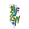
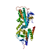
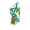
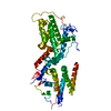
| Entry | Database: PDB / ID: 8uf2 | ||||||
|---|---|---|---|---|---|---|---|
| Title | Apo SOS2 crystal structure in P1 space group | ||||||
 Components Components | Son of sevenless homolog 2 | ||||||
 Keywords Keywords |  SIGNALING PROTEIN / SOS2 / Nucleotide Exchange Factor / GEF / KRAS / RAS / Fragment screening / FBLD SIGNALING PROTEIN / SOS2 / Nucleotide Exchange Factor / GEF / KRAS / RAS / Fragment screening / FBLD | ||||||
| Function / homology |  Function and homology information Function and homology informationregulation of pro-B cell differentiation / regulation of T cell differentiation in thymus / positive regulation of small GTPase mediated signal transduction / Interleukin-15 signaling / Activation of RAC1 / NRAGE signals death through JNK / regulation of T cell proliferation / B cell homeostasis / RAC1 GTPase cycle / guanyl-nucleotide exchange factor activity ...regulation of pro-B cell differentiation / regulation of T cell differentiation in thymus / positive regulation of small GTPase mediated signal transduction / Interleukin-15 signaling / Activation of RAC1 / NRAGE signals death through JNK / regulation of T cell proliferation / B cell homeostasis / RAC1 GTPase cycle / guanyl-nucleotide exchange factor activity / G alpha (12/13) signalling events / insulin receptor signaling pathway / Ras protein signal transduction / protein heterodimerization activity / plasma membrane / cytosolSimilarity search - Function | ||||||
| Biological species |  Homo sapiens (human) Homo sapiens (human) | ||||||
| Method | X-RAY DIFFRACTION / SYNCHROTRON / MOLECULAR REPLACEMENT / Resolution: 1.6 Å | ||||||
 Authors Authors | Gunn, R.J. / Lawson, J.D. | ||||||
| Funding support |  United States, 1items United States, 1items
| ||||||
 Citation Citation | Journal: J.Med.Chem. / Year: 2024 Title: Discovery of Five SOS2 Fragment Hits with Binding Modes Determined by SOS2 X-Ray Cocrystallography. Authors: Smith, C.R. / Chen, D. / Christensen, J.G. / Coulombe, R. / Fethiere, J. / Gunn, R.J. / Hollander, J. / Jones, B. / Ketcham, J.M. / Khare, S. / Kuehler, J. / Lawson, J.D. / Marx, M.A. / ...Authors: Smith, C.R. / Chen, D. / Christensen, J.G. / Coulombe, R. / Fethiere, J. / Gunn, R.J. / Hollander, J. / Jones, B. / Ketcham, J.M. / Khare, S. / Kuehler, J. / Lawson, J.D. / Marx, M.A. / Olson, P. / Pearson, K.E. / Ren, C. / Tsagris, D. / Ulaganathan, T. / Van't Veer, I. / Wang, X. / Ivetac, A. | ||||||
| History |
|
- Structure visualization
| Structure viewer | Molecule: MolmilJmol/JSmol |
|---|
- Downloads & links
-Download
| PDBx/mmCIF format | 8uf2.cif.gz | 249 KB | Display | PDBx/mmCIF format |
|---|---|---|---|---|
| PDB format | pdb8uf2.ent.gz | 161.4 KB | Display | PDB format |
| PDBx/mmJSON format | 8uf2.json.gz | Tree view | PDBx/mmJSON format | |
| Others |  Other downloads Other downloads |
-Validation report
| Arichive directory | https://data.pdbj.org/pub/pdb/validation_reports/uf/8uf2ftp://data.pdbj.org/pub/pdb/validation_reports/uf/8uf2 | HTTPS FTP |
|---|
-Related structure data

-Links
 PDBj
PDBj


- Assembly
| Deposited unit | 
| ||||||||||||
|---|---|---|---|---|---|---|---|---|---|---|---|---|---|
| 1 |
| ||||||||||||
| Unit cell |
|
-Components
| #1: Protein | Mass: 57312.637 Da / Num. of mol.: 1 Source method: isolated from a genetically manipulated source Source: (gene. exp.) Homo sapiens (human) / Gene: SOS2 / Production host:  Escherichia coli (E. coli) / References: UniProt: Q07890 Escherichia coli (E. coli) / References: UniProt: Q07890 |
|---|---|
| #2: Chemical | ChemComp-SO4 / Sulfate  Mass: 96.063 Da / Num. of mol.: 1 / Source method: obtained synthetically / Formula: SO4 Mass: 96.063 Da / Num. of mol.: 1 / Source method: obtained synthetically / Formula: SO4 |
| #3: Water | ChemComp-HOH / Water Mass: 18.015 Da / Num. of mol.: 649 / Source method: isolated from a natural source / Formula: H2O Mass: 18.015 Da / Num. of mol.: 649 / Source method: isolated from a natural source / Formula: H2O |
| Has ligand of interest | N |
-Experimental details
-Experiment
| Experiment | Method: X-RAY DIFFRACTION / Number of used crystals: 1 |
|---|
- Sample preparation
| Crystal | Density Matthews: 2.47 Å3/Da / Density % sol: 50.16 % |
|---|---|
| Crystal grow | Temperature: 281 K / Method: vapor diffusion, hanging drop / Details: 10% PEG 3350 |
-Data collection
| Diffraction | Mean temperature: 100 K / Serial crystal experiment: N |
|---|---|
| Diffraction source | Source: SYNCHROTRON / Site: APS / Beamline: 24-ID-E / Wavelength: 0.9796 Å |
| Detector | Type: DECTRIS EIGER X 16M / Detector: PIXEL / Date: Nov 11, 2021 |
| Radiation | Protocol: SINGLE WAVELENGTH / Monochromatic (M) / Laue (L): M / Scattering type: x-ray |
| Radiation wavelength | Wavelength: 0.9796 Å / Relative weight: 1 |
| Reflection | Resolution: 1.6→50 Å / Num. obs: 53261 / % possible obs: 73.65 % / Redundancy: 2.3 % / Biso Wilson estimate: 13.01 Å2 / CC1/2: 0.99 / Net I/σ(I): 19.4 |
| Reflection shell | Resolution: 1.6→1.63 Å / Num. unique obs: 576 / CC1/2: 0.67 |
- Processing
| Software |
| |||||||||||||||||||||||||||||||||||||||||||||||||||||||||||||||||||||||||||
|---|---|---|---|---|---|---|---|---|---|---|---|---|---|---|---|---|---|---|---|---|---|---|---|---|---|---|---|---|---|---|---|---|---|---|---|---|---|---|---|---|---|---|---|---|---|---|---|---|---|---|---|---|---|---|---|---|---|---|---|---|---|---|---|---|---|---|---|---|---|---|---|---|---|---|---|---|
| Refinement | Method to determine structure: MOLECULAR REPLACEMENT / Resolution: 1.6→44.09 Å / SU ML: 0.1812 / Cross valid method: FREE R-VALUE / σ(F): 2 / Phase error: 27.3479 Stereochemistry target values: GeoStd + Monomer Library + CDL v1.2
| |||||||||||||||||||||||||||||||||||||||||||||||||||||||||||||||||||||||||||
| Solvent computation | Shrinkage radii: 0.9 Å / VDW probe radii: 1.11 Å / Solvent model: FLAT BULK SOLVENT MODEL | |||||||||||||||||||||||||||||||||||||||||||||||||||||||||||||||||||||||||||
| Displacement parameters | Biso mean: 20.86 Å2 | |||||||||||||||||||||||||||||||||||||||||||||||||||||||||||||||||||||||||||
| Refinement step | Cycle: LAST / Resolution: 1.6→44.09 Å
| |||||||||||||||||||||||||||||||||||||||||||||||||||||||||||||||||||||||||||
| Refine LS restraints |
| |||||||||||||||||||||||||||||||||||||||||||||||||||||||||||||||||||||||||||
| LS refinement shell |
| |||||||||||||||||||||||||||||||||||||||||||||||||||||||||||||||||||||||||||
| Refinement TLS params. | Method: refined / Refine-ID: X-RAY DIFFRACTION
| |||||||||||||||||||||||||||||||||||||||||||||||||||||||||||||||||||||||||||
| Refinement TLS group | Refine-ID: X-RAY DIFFRACTION / Auth asym-ID: A / Label asym-ID: A
|
