 Movie
Movie Controller


+ Open data

- Basic information
| Entry | Database: PDB / ID: 8uch | ||||||
|---|---|---|---|---|---|---|---|
| Title | Thermophilic RNA Ligase from Palaeococcus pacificus K92A + ATP | ||||||
 Components Components | ATP dependent DNA ligase | ||||||
 Keywords Keywords |  LIGASE / RNA ligase / thermophilic / Archaea / Rnl3 / nucleotidyl-transferase LIGASE / RNA ligase / thermophilic / Archaea / Rnl3 / nucleotidyl-transferase | ||||||
| Function / homology | RNA ligase, Pab1020 family / RNA ligase Pab1020, C-terminal domain / Ligase Pab1020 C-terminal region / RNA ligase domain, REL/Rln2 / RNA ligase / ligase activity / ADENOSINE-5'-TRIPHOSPHATE / SPERMIDINE / ATP dependent DNA ligase Function and homology information Function and homology information | ||||||
| Biological species |  Palaeococcus pacificus DY20341 (archaea) Palaeococcus pacificus DY20341 (archaea) | ||||||
| Method | X-RAY DIFFRACTION / SYNCHROTRON / MOLECULAR REPLACEMENT / Resolution: 2.14 Å | ||||||
 Authors Authors | Rousseau, M.D. / Hicks, J.L. / Oulavallickal, T. / Williamson, A. / Arcus, V.L. / Patrick, M.W. | ||||||
| Funding support |  New Zealand, 1items New Zealand, 1items
| ||||||
 Citation Citation | Journal: Nucleic Acids Res. / Year: 2024 Title: Characterisation and engineering of a thermophilic RNA ligase from Palaeococcus pacificus. Authors: Rousseau, M. / Oulavallickal, T. / Williamson, A. / Arcus, V. / Patrick, W.M. / Hicks, J. | ||||||
| History |
|
- Structure visualization
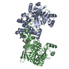
| Structure viewer | Molecule: MolmilJmol/JSmol |
|---|
- Downloads & links
-Download
| PDBx/mmCIF format | 8uch.cif.gz | 214.8 KB | Display | PDBx/mmCIF format |
|---|---|---|---|---|
| PDB format | pdb8uch.ent.gz | 139.9 KB | Display | PDB format |
| PDBx/mmJSON format | 8uch.json.gz | Tree view | PDBx/mmJSON format | |
| Others |  Other downloads Other downloads |
-Validation report
| Arichive directory | https://data.pdbj.org/pub/pdb/validation_reports/uc/8uchftp://data.pdbj.org/pub/pdb/validation_reports/uc/8uch | HTTPS FTP |
|---|
-Related structure data


-Links
 PDBj
PDBj
- Assembly
| Deposited unit | 
| ||||||||||||
|---|---|---|---|---|---|---|---|---|---|---|---|---|---|
| 1 | 
| ||||||||||||
| Unit cell |
| ||||||||||||
| Components on special symmetry positions |
|
-Components
-Protein , 1 types, 1 molecules A
| #1: Protein | Mass: 46916.027 Da / Num. of mol.: 1 / Mutation: K92A Source method: isolated from a genetically manipulated source Source: (gene. exp.) Palaeococcus pacificus DY20341 (archaea)Gene: PAP_02190 / Plasmid: PET28b / Production host:  Escherichia coli BL21(DE3) (bacteria) / References: UniProt: A0A075LQ94, RNA ligase (ATP) Escherichia coli BL21(DE3) (bacteria) / References: UniProt: A0A075LQ94, RNA ligase (ATP) |
|---|
-Non-polymers , 6 types, 224 molecules 


| #2: Chemical | ChemComp-ATP / Adenosine triphosphate Mass: 507.181 Da / Num. of mol.: 1 / Source method: obtained synthetically / Formula: C10H16N5O13P3 / Feature type: SUBJECT OF INVESTIGATION / Comment: ATP, energy-carrying molecule*YM Mass: 507.181 Da / Num. of mol.: 1 / Source method: obtained synthetically / Formula: C10H16N5O13P3 / Feature type: SUBJECT OF INVESTIGATION / Comment: ATP, energy-carrying molecule*YM | ||||||||
|---|---|---|---|---|---|---|---|---|---|
| #3: Chemical | ChemComp-GOL / Glycerol Mass: 92.094 Da / Num. of mol.: 4 / Source method: obtained synthetically / Formula: C3H8O3 Mass: 92.094 Da / Num. of mol.: 4 / Source method: obtained synthetically / Formula: C3H8O3#4: Chemical | ChemComp-SPD / | Spermidine Mass: 145.246 Da / Num. of mol.: 1 / Source method: obtained synthetically / Formula: C7H19N3 / Feature type: SUBJECT OF INVESTIGATION Mass: 145.246 Da / Num. of mol.: 1 / Source method: obtained synthetically / Formula: C7H19N3 / Feature type: SUBJECT OF INVESTIGATION#5: Chemical |  Mass: 24.305 Da / Num. of mol.: 3 / Source method: obtained synthetically / Formula: Mg / Feature type: SUBJECT OF INVESTIGATION Mass: 24.305 Da / Num. of mol.: 3 / Source method: obtained synthetically / Formula: Mg / Feature type: SUBJECT OF INVESTIGATION#6: Chemical |  Mass: 22.990 Da / Num. of mol.: 3 / Source method: obtained synthetically / Formula: Na Mass: 22.990 Da / Num. of mol.: 3 / Source method: obtained synthetically / Formula: Na#7: Water | ChemComp-HOH / | WaterMass: 18.015 Da / Num. of mol.: 212 / Source method: isolated from a natural source / Formula: H2O |
-Details
| Has ligand of interest | Y |
|---|
-Experimental details
-Experiment
| Experiment | Method: X-RAY DIFFRACTION / Number of used crystals: 1 |
|---|
- Sample preparation
| Crystal | Density Matthews: 2.98 Å3/Da / Density % sol: 58.69 % |
|---|---|
| Crystal grow | Temperature: 291.15 K / Method: vapor diffusion, hanging drop / pH: 6.5 Details: 80 mM strontium chloride hexahydrate, 20 mM magnesium chloride hexahydrate, 40 mM sodium cacodylate trihydrate pH 7.0, 20% v/v (+/-)-2-methyl-2,4-pentanediol, 12 mM spermine tetrahydrochloride |
-Data collection
| Diffraction | Mean temperature: 100 K / Ambient temp details: Liquid nitrogen / Serial crystal experiment: N |
|---|---|
| Diffraction source | Source: SYNCHROTRON / Site: Australian Synchrotron  / Beamline: MX2 / Wavelength: 0.953659 Å / Beamline: MX2 / Wavelength: 0.953659 Å |
| Detector | Type: DECTRIS EIGER X 16M / Detector: PIXEL / Date: Mar 11, 2022 |
| Radiation | Protocol: SINGLE WAVELENGTH / Monochromatic (M) / Laue (L): M / Scattering type: x-ray |
| Radiation wavelength | Wavelength: 0.953659 Å / Relative weight: 1 |
| Reflection | Resolution: 2.14→46.72 Å / Num. obs: 31266 / % possible obs: 99.97 % / Redundancy: 13.3 % / Biso Wilson estimate: 21.41 Å2 / Rmerge(I) obs: 0.1088 / Net I/σ(I): 16.75 |
| Reflection shell | Resolution: 2.217→46.72 Å / Rmerge(I) obs: 0.9675 / Mean I/σ(I) obs: 2.55 / Num. unique obs: 3098 |
- Processing
| Software |
| |||||||||||||||||||||||||||||||||||||||||||||||||||||||||||||||||||||||||||||||||||||||||||||||||||||||||
|---|---|---|---|---|---|---|---|---|---|---|---|---|---|---|---|---|---|---|---|---|---|---|---|---|---|---|---|---|---|---|---|---|---|---|---|---|---|---|---|---|---|---|---|---|---|---|---|---|---|---|---|---|---|---|---|---|---|---|---|---|---|---|---|---|---|---|---|---|---|---|---|---|---|---|---|---|---|---|---|---|---|---|---|---|---|---|---|---|---|---|---|---|---|---|---|---|---|---|---|---|---|---|---|---|---|---|
| Refinement | Method to determine structure: MOLECULAR REPLACEMENT / Resolution: 2.14→46.72 Å / SU ML: 0.1858 / Cross valid method: FREE R-VALUE / σ(F): 1.39 / Phase error: 19.23 Stereochemistry target values: GeoStd + Monomer Library + CDL v1.2
| |||||||||||||||||||||||||||||||||||||||||||||||||||||||||||||||||||||||||||||||||||||||||||||||||||||||||
| Solvent computation | Shrinkage radii: 0.9 Å / VDW probe radii: 1.1 Å / Solvent model: FLAT BULK SOLVENT MODEL | |||||||||||||||||||||||||||||||||||||||||||||||||||||||||||||||||||||||||||||||||||||||||||||||||||||||||
| Displacement parameters | Biso mean: 27.55 Å2 | |||||||||||||||||||||||||||||||||||||||||||||||||||||||||||||||||||||||||||||||||||||||||||||||||||||||||
| Refinement step | Cycle: LAST / Resolution: 2.14→46.72 Å
| |||||||||||||||||||||||||||||||||||||||||||||||||||||||||||||||||||||||||||||||||||||||||||||||||||||||||
| Refine LS restraints |
| |||||||||||||||||||||||||||||||||||||||||||||||||||||||||||||||||||||||||||||||||||||||||||||||||||||||||
| LS refinement shell |
| |||||||||||||||||||||||||||||||||||||||||||||||||||||||||||||||||||||||||||||||||||||||||||||||||||||||||
| Refinement TLS params. | Method: refined / Origin x: 57.7905638155 Å / Origin y: 20.7023073719 Å / Origin z: 48.3891654255 Å
| |||||||||||||||||||||||||||||||||||||||||||||||||||||||||||||||||||||||||||||||||||||||||||||||||||||||||
| Refinement TLS group | Selection details: all |
