 Movie
Movie Controller


+ Open data

- Basic information
| Entry | Database: PDB / ID: 8u8p | ||||||
|---|---|---|---|---|---|---|---|
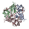
| Title | S292F Streptomyces coelicolor Laccase | ||||||
 Components Components | Copper oxidase | ||||||
 Keywords Keywords |  OXIDOREDUCTASE / Laccase / Copper OXIDOREDUCTASE / Laccase / Copper | ||||||
| Function / homology |  Function and homology information Function and homology information | ||||||
| Biological species |  Streptomyces coelicolor (bacteria) Streptomyces coelicolor (bacteria) | ||||||
| Method | X-RAY DIFFRACTION / SYNCHROTRON / MOLECULAR REPLACEMENT / Resolution: 2.2 Å | ||||||
 Authors Authors | Wang, J.-X. / Lu, Y. | ||||||
| Funding support |  United States, 1items United States, 1items
| ||||||
 Citation Citation | Journal: Angew.Chem.Int.Ed.Engl. / Year: 2023 Title: Increasing Reduction Potentials of Type 1 Copper Center and Catalytic Efficiency of Small Laccase from Streptomyces coelicolor through Secondary Coordination Sphere Mutations. Authors: Wang, J.X. / Vilbert, A.C. / Cui, C. / Mirts, E.N. / Williams, L.H. / Kim, W. / Jessie Zhang, Y. / Lu, Y. | ||||||
| History |
|
- Structure visualization
| Structure viewer | Molecule: MolmilJmol/JSmol |
|---|
- Downloads & links
-Download
| PDBx/mmCIF format | 8u8p.cif.gz | 240.7 KB | Display | PDBx/mmCIF format |
|---|---|---|---|---|
| PDB format | pdb8u8p.ent.gz | 155.9 KB | Display | PDB format |
| PDBx/mmJSON format | 8u8p.json.gz | Tree view | PDBx/mmJSON format | |
| Others |  Other downloads Other downloads |
-Validation report
| Arichive directory | https://data.pdbj.org/pub/pdb/validation_reports/u8/8u8pftp://data.pdbj.org/pub/pdb/validation_reports/u8/8u8p | HTTPS FTP |
|---|
-Related structure data


-Links
 PDBj
PDBj


- Assembly
| Deposited unit | 
| ||||||||||||
|---|---|---|---|---|---|---|---|---|---|---|---|---|---|
| 1 |
| ||||||||||||
| Unit cell |
|
-Components
-Protein , 1 types, 3 molecules ABC
| #1: Protein | Mass: 38063.242 Da / Num. of mol.: 3 / Mutation: S292F Source method: isolated from a genetically manipulated source Source: (gene. exp.) Streptomyces coelicolor (bacteria) / Gene: SCO6712 / Production host: Escherichia coli (E. coli) / References: UniProt: Q9XAL8 |
|---|
-Non-polymers , 8 types, 473 molecules 


| #2: Chemical | ChemComp-CU / Copper Mass: 63.546 Da / Num. of mol.: 12 / Source method: isolated from a natural source / Formula: Cu / Feature type: SUBJECT OF INVESTIGATION Mass: 63.546 Da / Num. of mol.: 12 / Source method: isolated from a natural source / Formula: Cu / Feature type: SUBJECT OF INVESTIGATION#3: Chemical | Hydroxide Mass: 17.007 Da / Num. of mol.: 3 / Source method: isolated from a natural source / Formula: HO / Feature type: SUBJECT OF INVESTIGATION Mass: 17.007 Da / Num. of mol.: 3 / Source method: isolated from a natural source / Formula: HO / Feature type: SUBJECT OF INVESTIGATION#4: Chemical | ChemComp-GLY / Glycine Type: peptide linking / Mass: 75.067 Da / Num. of mol.: 15 / Source method: obtained synthetically / Formula: C2H5NO2 Type: peptide linking / Mass: 75.067 Da / Num. of mol.: 15 / Source method: obtained synthetically / Formula: C2H5NO2#5: Chemical | Diethylene glycol Mass: 106.120 Da / Num. of mol.: 3 / Source method: obtained synthetically / Formula: C4H10O3 Mass: 106.120 Da / Num. of mol.: 3 / Source method: obtained synthetically / Formula: C4H10O3#6: Chemical | Polyethylene glycol Mass: 150.173 Da / Num. of mol.: 3 / Source method: obtained synthetically / Formula: C6H14O4 Mass: 150.173 Da / Num. of mol.: 3 / Source method: obtained synthetically / Formula: C6H14O4#7: Chemical | ChemComp-PG4 / Polyethylene glycol Mass: 194.226 Da / Num. of mol.: 6 / Source method: obtained synthetically / Formula: C8H18O5 / Comment: precipitant*YM Mass: 194.226 Da / Num. of mol.: 6 / Source method: obtained synthetically / Formula: C8H18O5 / Comment: precipitant*YM#8: Chemical | ChemComp-BO3 / | Boric acid Mass: 61.833 Da / Num. of mol.: 1 / Source method: obtained synthetically / Formula: BH3O3 Mass: 61.833 Da / Num. of mol.: 1 / Source method: obtained synthetically / Formula: BH3O3#9: Water | ChemComp-HOH / | WaterMass: 18.015 Da / Num. of mol.: 430 / Source method: isolated from a natural source / Formula: H2O |
|---|
-Details
| Has ligand of interest | Y |
|---|
-Experimental details
-Experiment
| Experiment | Method: X-RAY DIFFRACTION / Number of used crystals: 1 |
|---|
- Sample preparation
| Crystal grow | Temperature: 296 K / Method: vapor diffusion, hanging drop / pH: 9 Details: Crystals were prepared using hanging drop vapor-diffusion technique at room temperature (~296 K). Protein is at a concentration of 18.5 mg/ml in 50 mM H3BO3, 0.1 M NaCl, pH 9.0 buffer. The ...Details: Crystals were prepared using hanging drop vapor-diffusion technique at room temperature (~296 K). Protein is at a concentration of 18.5 mg/ml in 50 mM H3BO3, 0.1 M NaCl, pH 9.0 buffer. The well buffer contains 0.1 M glycine, 0.3-0.6 M NaCl, pH 9.0, and 37-39% (v/v) PEG (polyethylene glycol) monomethyl ether 550. 500 uL of well buffer is added to each well and protein is mixed with well buffer at a 1.5 uL:1.5 uL ratio. The crystal growth time was ca. 1-2 weeks |
|---|
-Data collection
| Diffraction | Mean temperature: 100 K / Serial crystal experiment: N |
|---|---|
| Diffraction source | Source: SYNCHROTRON / Site: ALS / Beamline: 5.0.2 / Wavelength: 1.00003 Å |
| Detector | Type: DECTRIS PILATUS3 6M / Detector: PIXEL / Date: Dec 18, 2022 |
| Radiation | Protocol: SINGLE WAVELENGTH / Monochromatic (M) / Laue (L): M / Scattering type: x-ray |
| Radiation wavelength | Wavelength: 1.00003 Å / Relative weight: 1 |
| Reflection | Resolution: 2.2→62.69 Å / Num. obs: 142572 / % possible obs: 99.97 % / Redundancy: 13.4 % / Biso Wilson estimate: 38.98 Å2 / CC1/2: 0.998 / Rmerge(I) obs: 0.1663 / Net I/σ(I): 11.31 |
| Reflection shell | Resolution: 2.2→2.279 Å / Rmerge(I) obs: 1.525 / Num. unique obs: 14126 / CC1/2: 0.754 |
- Processing
| Software |
| |||||||||||||||||||||||||||||||||||||||||||||||||||||||||||||||||||||||||||||||||||||||||||||||||||||||||||||||||||||||||||||||||||||||||||||||||||||||||||||||||||||||||||||||||||||||||||||||||||||||||||||||||||||||||
|---|---|---|---|---|---|---|---|---|---|---|---|---|---|---|---|---|---|---|---|---|---|---|---|---|---|---|---|---|---|---|---|---|---|---|---|---|---|---|---|---|---|---|---|---|---|---|---|---|---|---|---|---|---|---|---|---|---|---|---|---|---|---|---|---|---|---|---|---|---|---|---|---|---|---|---|---|---|---|---|---|---|---|---|---|---|---|---|---|---|---|---|---|---|---|---|---|---|---|---|---|---|---|---|---|---|---|---|---|---|---|---|---|---|---|---|---|---|---|---|---|---|---|---|---|---|---|---|---|---|---|---|---|---|---|---|---|---|---|---|---|---|---|---|---|---|---|---|---|---|---|---|---|---|---|---|---|---|---|---|---|---|---|---|---|---|---|---|---|---|---|---|---|---|---|---|---|---|---|---|---|---|---|---|---|---|---|---|---|---|---|---|---|---|---|---|---|---|---|---|---|---|---|---|---|---|---|---|---|---|---|---|---|---|---|---|---|---|---|
| Refinement | Method to determine structure: MOLECULAR REPLACEMENT / Resolution: 2.2→62.69 Å / SU ML: 0.202 / Cross valid method: FREE R-VALUE / σ(F): 1.35 / Phase error: 16.7106 Stereochemistry target values: GeoStd + Monomer Library + CDL v1.2
| |||||||||||||||||||||||||||||||||||||||||||||||||||||||||||||||||||||||||||||||||||||||||||||||||||||||||||||||||||||||||||||||||||||||||||||||||||||||||||||||||||||||||||||||||||||||||||||||||||||||||||||||||||||||||
| Solvent computation | Shrinkage radii: 0.9 Å / VDW probe radii: 1.1 Å / Solvent model: FLAT BULK SOLVENT MODEL | |||||||||||||||||||||||||||||||||||||||||||||||||||||||||||||||||||||||||||||||||||||||||||||||||||||||||||||||||||||||||||||||||||||||||||||||||||||||||||||||||||||||||||||||||||||||||||||||||||||||||||||||||||||||||
| Displacement parameters | Biso mean: 41.41 Å2 | |||||||||||||||||||||||||||||||||||||||||||||||||||||||||||||||||||||||||||||||||||||||||||||||||||||||||||||||||||||||||||||||||||||||||||||||||||||||||||||||||||||||||||||||||||||||||||||||||||||||||||||||||||||||||
| Refinement step | Cycle: LAST / Resolution: 2.2→62.69 Å
| |||||||||||||||||||||||||||||||||||||||||||||||||||||||||||||||||||||||||||||||||||||||||||||||||||||||||||||||||||||||||||||||||||||||||||||||||||||||||||||||||||||||||||||||||||||||||||||||||||||||||||||||||||||||||
| Refine LS restraints |
| |||||||||||||||||||||||||||||||||||||||||||||||||||||||||||||||||||||||||||||||||||||||||||||||||||||||||||||||||||||||||||||||||||||||||||||||||||||||||||||||||||||||||||||||||||||||||||||||||||||||||||||||||||||||||
| LS refinement shell |
|
