 Movie
Movie Controller

[English] 日本語
 Yorodumi
- PDB-8u7i: Structure of the phage immune evasion protein Gad1 bound to the G... -
+ Open data

- Basic information
| Entry | Database: PDB / ID: 8u7i | ||||||||||||||||||
|---|---|---|---|---|---|---|---|---|---|---|---|---|---|---|---|---|---|---|---|
| Title | Structure of the phage immune evasion protein Gad1 bound to the Gabija GajAB complex | ||||||||||||||||||
 Components Components |
| ||||||||||||||||||
 Keywords Keywords |  VIRAL PROTEIN / viral immune evasion / phage / bacteria / anti-phage defense complex / DNA nuclease / DNA helicase VIRAL PROTEIN / viral immune evasion / phage / bacteria / anti-phage defense complex / DNA nuclease / DNA helicase | ||||||||||||||||||
| Function / homology |  Function and homology informationDNA helicase activity / defense response to virus / endonuclease activity / Hydrolases; Acting on ester bonds / hydrolase activity / DNA binding / ATP binding / metal ion binding Function and homology informationDNA helicase activity / defense response to virus / endonuclease activity / Hydrolases; Acting on ester bonds / hydrolase activity / DNA binding / ATP binding / metal ion bindingSimilarity search - Function | ||||||||||||||||||
| Biological species |   Bacillus phage phi3T (virus) Bacillus phage phi3T (virus) | ||||||||||||||||||
| Method | ELECTRON MICROSCOPY / single particle reconstruction / cryo EM / Resolution: 2.57 Å | ||||||||||||||||||
 Authors Authors | Antine, S.P. / Johnson, A.G. / Mooney, S.E. / Mayer, M.L. / Kranzsuch, P.J. | ||||||||||||||||||
| Funding support |  United States, 5items United States, 5items
| ||||||||||||||||||
 Citation Citation | Journal: Nature / Year: 2024 Title: Structural basis of Gabija anti-phage defence and viral immune evasion. Authors: Sadie P Antine / Alex G Johnson / Sarah E Mooney / Azita Leavitt / Megan L Mayer / Erez Yirmiya / Gil Amitai / Rotem Sorek / Philip J Kranzusch /  Abstract: Bacteria encode hundreds of diverse defence systems that protect them from viral infection and inhibit phage propagation. Gabija is one of the most prevalent anti-phage defence systems, occurring in ...Bacteria encode hundreds of diverse defence systems that protect them from viral infection and inhibit phage propagation. Gabija is one of the most prevalent anti-phage defence systems, occurring in more than 15% of all sequenced bacterial and archaeal genomes, but the molecular basis of how Gabija defends cells from viral infection remains poorly understood. Here we use X-ray crystallography and cryo-electron microscopy (cryo-EM) to define how Gabija proteins assemble into a supramolecular complex of around 500 kDa that degrades phage DNA. Gabija protein A (GajA) is a DNA endonuclease that tetramerizes to form the core of the anti-phage defence complex. Two sets of Gabija protein B (GajB) dimers dock at opposite sides of the complex and create a 4:4 GajA-GajB assembly (hereafter, GajAB) that is essential for phage resistance in vivo. We show that a phage-encoded protein, Gabija anti-defence 1 (Gad1), directly binds to the Gabija GajAB complex and inactivates defence. A cryo-EM structure of the virally inhibited state shows that Gad1 forms an octameric web that encases the GajAB complex and inhibits DNA recognition and cleavage. Our results reveal the structural basis of assembly of the Gabija anti-phage defence complex and define a unique mechanism of viral immune evasion. #1: Journal: To Be PublishedTitle: Phages overcome bacterial immunity via diverse anti-defense proteins Authors: Yirmiya, E. / Leavitt, A. / Lu, A. / Avraham, C. / Osterman, I. / Garb, J. / Antine, S.P. / Mooney, S.E. / Hobbs, S.J. / Kranzusch, P.J. / Amitai, G. / Sorek, R. | ||||||||||||||||||
| History |
|
- Structure visualization
| Structure viewer | Molecule: MolmilJmol/JSmol |
|---|
- Downloads & links
-Download
| PDBx/mmCIF format | 8u7i.cif.gz | 1.2 MB | Display | PDBx/mmCIF format |
|---|---|---|---|---|
| PDB format | pdb8u7i.ent.gz | 785.1 KB | Display | PDB format |
| PDBx/mmJSON format | 8u7i.json.gz | Tree view | PDBx/mmJSON format | |
| Others |  Other downloads Other downloads |
-Validation report
| Arichive directory | https://data.pdbj.org/pub/pdb/validation_reports/u7/8u7iftp://data.pdbj.org/pub/pdb/validation_reports/u7/8u7i | HTTPS FTP |
|---|
-Related structure data
| Related structure data | 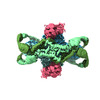 41983MC  8sm3C M: map data used to model this data C: citing same article ( |
|---|---|
| Similar structure data |
-Links
 PDBj
PDBj

- Assembly
| Deposited unit | 
|
|---|---|
| 1 |
|
-Components
| #1: Protein | Mass: 78045.656 Da / Num. of mol.: 4 Source method: isolated from a genetically manipulated source Source: (gene. exp.) Escherichia coli BL21(DE3) (bacteria) / References: UniProt: J8H9C1#2: Protein | Mass: 57139.992 Da / Num. of mol.: 4 Source method: isolated from a genetically manipulated source Source: (gene. exp.) Escherichia coli BL21(DE3) (bacteria) / References: UniProt: J8HQ06#3: Protein | Mass: 34919.184 Da / Num. of mol.: 8 Source method: isolated from a genetically manipulated source Source: (gene. exp.) Bacillus phage phi3T (virus) / Gene: phi3T_128 / Production host: Escherichia coli BL21(DE3) (bacteria) / References: UniProt: A0A1P8CWZ3 |
|---|
-Experimental details
-Experiment
| Experiment | Method: ELECTRON MICROSCOPY |
|---|---|
| EM experiment | Aggregation state: 3D ARRAY / 3D reconstruction method: single particle reconstruction |
- Sample preparation
| Component |
| ||||||||||||||||||||||||
|---|---|---|---|---|---|---|---|---|---|---|---|---|---|---|---|---|---|---|---|---|---|---|---|---|---|
| Molecular weight |
| ||||||||||||||||||||||||
| Source (natural) |
| ||||||||||||||||||||||||
| Source (recombinant) |
| ||||||||||||||||||||||||
| Buffer solution | pH: 7.5 Details: 20 mM HEPES-KOH pH 7.5, 20 mM KCl, and 1 mM TCEP-KOH | ||||||||||||||||||||||||
| Specimen | Conc.: 1 mg/ml / Embedding applied: NO / Shadowing applied: NO / Staining applied: NO / Vitrification applied: YES | ||||||||||||||||||||||||
| Vitrification | Instrument: FEI VITROBOT MARK IV / Cryogen name: ETHANE / Humidity: 100 % / Chamber temperature: 277.15 K |
- Electron microscopy imaging
| Experimental equipment |  Model: Titan Krios / Image courtesy: FEI Company |
|---|---|
| Microscopy | Model: FEI TITAN KRIOS |
| Electron gun | Electron source: FIELD EMISSION GUN / Accelerating voltage: 300 kV / Illumination mode: FLOOD BEAM |
| Electron lens | Mode: BRIGHT FIELDBright-field microscopy / Nominal defocus max: 1900 nm / Nominal defocus min: 800 nm |
| Image recording | Electron dose: 41.1 e/Å2 / Film or detector model: GATAN K3 (6k x 4k) |
- Processing
| CTF correction | Type: NONE | ||||||||||||||||||||||||
|---|---|---|---|---|---|---|---|---|---|---|---|---|---|---|---|---|---|---|---|---|---|---|---|---|---|
| 3D reconstruction | Resolution: 2.57 Å / Resolution method: FSC 0.143 CUT-OFF / Num. of particles: 351193 / Symmetry type: POINT | ||||||||||||||||||||||||
| Refinement | Cross valid method: NONE Stereochemistry target values: GeoStd + Monomer Library + CDL v1.2 | ||||||||||||||||||||||||
| Displacement parameters | Biso mean: 170.99 Å2 | ||||||||||||||||||||||||
| Refine LS restraints |
|
