 Movie
Movie Controller

[English] 日本語
 Yorodumi
- PDB-8u6d: Crystal Structure of HIV-1 Reverse Transcriptase in Complex with ... -
+ Open data

- Basic information
| Entry | Database: PDB / ID: 8u6d | ||||||
|---|---|---|---|---|---|---|---|
| Title | Crystal Structure of HIV-1 Reverse Transcriptase in Complex with N-(2-(4-chloro-3-(3-chloro-5-cyanophenoxy)phenoxy)ethyl)-N-methylacrylamide (JLJ736), a non-nucleoside inhibitor | ||||||
 Components Components |
| ||||||
 Keywords Keywords |  VIRAL PROTEIN/Inhibitor / REVERSE TRANSCRIPTASE / ANTIVIRAL / DRUG DESIGN / HIV-1 / VIRAL PROTEIN / VIRAL PROTEIN-Inhibitor complex VIRAL PROTEIN/Inhibitor / REVERSE TRANSCRIPTASE / ANTIVIRAL / DRUG DESIGN / HIV-1 / VIRAL PROTEIN / VIRAL PROTEIN-Inhibitor complex | ||||||
| Function / homology |  Function and homology informationHIV-1 retropepsin / : / retroviral ribonuclease H / exoribonuclease H / : / exoribonuclease H activity / host multivesicular body / DNA integration / RNA-directed DNA polymerase / viral genome integration into host DNA ...HIV-1 retropepsin / : / retroviral ribonuclease H / exoribonuclease H / : / exoribonuclease H activity / host multivesicular body / DNA integration / RNA-directed DNA polymerase / viral genome integration into host DNA / viral penetration into host nucleus / establishment of integrated proviral latency / RNA-directed DNA polymerase activity / Transferases; Transferring phosphorus-containing groups; Nucleotidyltransferases / RNA-DNA hybrid ribonuclease activity / viral nucleocapsid / DNA recombination / Hydrolases; Acting on ester bonds / DNA-directed DNA polymerase / aspartic-type endopeptidase activity / DNA-directed DNA polymerase activity / symbiont entry into host cell / symbiont-mediated suppression of host gene expression / lipid binding / host cell nucleus / host cell plasma membrane / virion membrane / structural molecule activity / proteolysis / DNA binding / RNA binding / zinc ion binding / membrane Function and homology informationHIV-1 retropepsin / : / retroviral ribonuclease H / exoribonuclease H / : / exoribonuclease H activity / host multivesicular body / DNA integration / RNA-directed DNA polymerase / viral genome integration into host DNA ...HIV-1 retropepsin / : / retroviral ribonuclease H / exoribonuclease H / : / exoribonuclease H activity / host multivesicular body / DNA integration / RNA-directed DNA polymerase / viral genome integration into host DNA / viral penetration into host nucleus / establishment of integrated proviral latency / RNA-directed DNA polymerase activity / Transferases; Transferring phosphorus-containing groups; Nucleotidyltransferases / RNA-DNA hybrid ribonuclease activity / viral nucleocapsid / DNA recombination / Hydrolases; Acting on ester bonds / DNA-directed DNA polymerase / aspartic-type endopeptidase activity / DNA-directed DNA polymerase activity / symbiont entry into host cell / symbiont-mediated suppression of host gene expression / lipid binding / host cell nucleus / host cell plasma membrane / virion membrane / structural molecule activity / proteolysis / DNA binding / RNA binding / zinc ion binding / membraneSimilarity search - Function | ||||||
| Biological species |   Human immunodeficiency virus 1 Human immunodeficiency virus 1 | ||||||
| Method | X-RAY DIFFRACTION / SYNCHROTRON / MOLECULAR REPLACEMENT / Resolution: 2.33 Å | ||||||
 Authors Authors | Hollander, K. / Carter, Z. / Jorgensen, W.L. / Anderson, K.S. | ||||||
| Funding support |  United States, 1items United States, 1items
| ||||||
 Citation Citation | Journal: Eur.J.Med.Chem. / Year: 2023 Title: Covalent and noncovalent strategies for targeting Lys102 in HIV-1 reverse transcriptase. Authors: Prucha, G.R. / Henry, S. / Hollander, K. / Carter, Z.J. / Spasov, K.A. / Jorgensen, W.L. / Anderson, K.S. | ||||||
| History |
|
- Structure visualization
| Structure viewer | Molecule: MolmilJmol/JSmol |
|---|
- Downloads & links
-Download
| PDBx/mmCIF format | 8u6d.cif.gz | 200.1 KB | Display | PDBx/mmCIF format |
|---|---|---|---|---|
| PDB format | pdb8u6d.ent.gz | 153.4 KB | Display | PDB format |
| PDBx/mmJSON format | 8u6d.json.gz | Tree view | PDBx/mmJSON format | |
| Others |  Other downloads Other downloads |
-Validation report
| Arichive directory | https://data.pdbj.org/pub/pdb/validation_reports/u6/8u6dftp://data.pdbj.org/pub/pdb/validation_reports/u6/8u6d | HTTPS FTP |
|---|
-Related structure data
| Related structure data |  8u69C  8u6aC 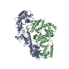 8u6bC  8u6cC 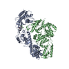 8u6eC  8u6fC 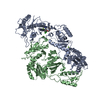 8u6gC 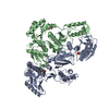 8u6hC 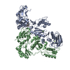 8u6iC 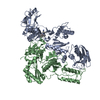 8u6jC  8u6kC  8u6lC 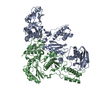 8u6mC  8u6nC  8u6oC  8u6pC 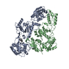 8u6qC  8u6rC  8u6sC  8u6tC C: citing same article ( |
|---|---|
| Similar structure data |
-Links
 PDBj
PDBj


- Assembly
| Deposited unit | 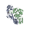
| ||||||||
|---|---|---|---|---|---|---|---|---|---|
| 1 |
| ||||||||
| Unit cell |
|
-Components
| #1: Protein | Mass: 63989.238 Da / Num. of mol.: 1 / Mutation: C879S, K172A, K173A Source method: isolated from a genetically manipulated source Source: (gene. exp.) Human immunodeficiency virus 1 / Production host:  Escherichia coli (E. coli) Escherichia coli (E. coli)References: UniProt: P03366, RNA-directed DNA polymerase, DNA-directed DNA polymerase, retroviral ribonuclease H |
|---|---|
| #2: Protein | Mass: 50039.488 Da / Num. of mol.: 1 / Mutation: C879S Source method: isolated from a genetically manipulated source Source: (gene. exp.) Human immunodeficiency virus 1 / Gene: gag-pol / Production host: Escherichia coli (E. coli) / References: UniProt: P03366 |
| #3: Chemical | ChemComp-VTS /   Mass: 391.248 Da / Num. of mol.: 1 / Source method: obtained synthetically / Formula: C19H16Cl2N2O3 / Feature type: SUBJECT OF INVESTIGATION Mass: 391.248 Da / Num. of mol.: 1 / Source method: obtained synthetically / Formula: C19H16Cl2N2O3 / Feature type: SUBJECT OF INVESTIGATION |
| #4: Water | ChemComp-HOH / Water Mass: 18.015 Da / Num. of mol.: 122 / Source method: isolated from a natural source / Formula: H2O Mass: 18.015 Da / Num. of mol.: 122 / Source method: isolated from a natural source / Formula: H2O |
| Has ligand of interest | Y |
-Experimental details
-Experiment
| Experiment | Method: X-RAY DIFFRACTION / Number of used crystals: 1 |
|---|
- Sample preparation
| Crystal | Density Matthews: 2.83 Å3/Da / Density % sol: 56.46 % |
|---|---|
| Crystal grow | Temperature: 277 K / Method: vapor diffusion, hanging drop / pH: 5 Details: 50 mM MES pH 5.0, 20% PEG 8000, 100 mM ammonium sulfate, 15 mM magnesium sulfate, and 5 mM spermine PH range: 5.0-7.5 |
-Data collection
| Diffraction | Mean temperature: 100 K / Serial crystal experiment: N |
|---|---|
| Diffraction source | Source: SYNCHROTRON / Site: NSLS-II / Beamline: 17-ID-1 / Wavelength: 0.9201 Å |
| Detector | Type: DECTRIS EIGER X 9M / Detector: PIXEL / Date: Jul 22, 2022 |
| Radiation | Protocol: SINGLE WAVELENGTH / Monochromatic (M) / Laue (L): M / Scattering type: x-ray |
| Radiation wavelength | Wavelength: 0.9201 Å / Relative weight: 1 |
| Reflection | Resolution: 2.33→80.06 Å / Num. obs: 54256 / % possible obs: 100 % / Redundancy: 7 % / CC1/2: 0.998 / Rmerge(I) obs: 0.106 / Rpim(I) all: 0.043 / Rrim(I) all: 0.115 / Χ2: 1 / Net I/σ(I): 10.8 / Num. measured all: 380742 |
| Reflection shell | Resolution: 2.33→2.46 Å / % possible obs: 100 % / Redundancy: 7.3 % / Rmerge(I) obs: 1.786 / Num. measured all: 57590 / Num. unique obs: 7868 / CC1/2: 0.488 / Rpim(I) all: 0.706 / Rrim(I) all: 1.922 / Χ2: 0.92 / Net I/σ(I) obs: 1.2 |
- Processing
| Software |
| ||||||||||||||||||||||||||||||||||||||||||||||||||||||||||||||||||||||||||||||||||||||||||||||||||||||||||||||||||||||||||||||||||||||||||||
|---|---|---|---|---|---|---|---|---|---|---|---|---|---|---|---|---|---|---|---|---|---|---|---|---|---|---|---|---|---|---|---|---|---|---|---|---|---|---|---|---|---|---|---|---|---|---|---|---|---|---|---|---|---|---|---|---|---|---|---|---|---|---|---|---|---|---|---|---|---|---|---|---|---|---|---|---|---|---|---|---|---|---|---|---|---|---|---|---|---|---|---|---|---|---|---|---|---|---|---|---|---|---|---|---|---|---|---|---|---|---|---|---|---|---|---|---|---|---|---|---|---|---|---|---|---|---|---|---|---|---|---|---|---|---|---|---|---|---|---|---|---|
| Refinement | Method to determine structure: MOLECULAR REPLACEMENT / Resolution: 2.33→35.22 Å / SU ML: 0.4 / Cross valid method: FREE R-VALUE / σ(F): 1.34 / Phase error: 29.87 / Stereochemistry target values: ML
| ||||||||||||||||||||||||||||||||||||||||||||||||||||||||||||||||||||||||||||||||||||||||||||||||||||||||||||||||||||||||||||||||||||||||||||
| Solvent computation | Shrinkage radii: 0.9 Å / VDW probe radii: 1.1 Å / Solvent model: FLAT BULK SOLVENT MODEL | ||||||||||||||||||||||||||||||||||||||||||||||||||||||||||||||||||||||||||||||||||||||||||||||||||||||||||||||||||||||||||||||||||||||||||||
| Refinement step | Cycle: LAST / Resolution: 2.33→35.22 Å
| ||||||||||||||||||||||||||||||||||||||||||||||||||||||||||||||||||||||||||||||||||||||||||||||||||||||||||||||||||||||||||||||||||||||||||||
| Refine LS restraints |
| ||||||||||||||||||||||||||||||||||||||||||||||||||||||||||||||||||||||||||||||||||||||||||||||||||||||||||||||||||||||||||||||||||||||||||||
| LS refinement shell |
|
