 Movie
Movie Controller


+ Open data

- Basic information
| Entry | Database: PDB / ID: 8tq8 | ||||||
|---|---|---|---|---|---|---|---|
| Title | Crystal structure of Fab.34.5.8 in complex with MHC-I (H2-Dd) | ||||||
 Components Components |
| ||||||
 Keywords Keywords |  IMMUNE SYSTEM / histocompatibility complex class I / MHC-I / immune response / immune system Fab / antibody / anti-MHC antibody / cancer tumor IMMUNE SYSTEM / histocompatibility complex class I / MHC-I / immune response / immune system Fab / antibody / anti-MHC antibody / cancer tumor | ||||||
| Function / homology |  Function and homology information Function and homology informationSynthesis and processing of ENV and VPU / evasion of host immune response / TAP1 binding / TAP2 binding / Alpha-defensins / positive regulation of antibody-dependent cellular cytotoxicity / regulation of natural killer cell mediated immunity / positive regulation of TRAIL production / antigen processing and presentation of exogenous peptide antigen via MHC class Ib / MHC class Ib protein complex ...Synthesis and processing of ENV and VPU / evasion of host immune response / TAP1 binding / TAP2 binding / Alpha-defensins / positive regulation of antibody-dependent cellular cytotoxicity / regulation of natural killer cell mediated immunity / positive regulation of TRAIL production / antigen processing and presentation of exogenous peptide antigen via MHC class Ib / MHC class Ib protein complex / positive regulation of natural killer cell mediated immunity / positive regulation of natural killer cell cytokine production / natural killer cell tolerance induction / natural killer cell lectin-like receptor binding / negative regulation of natural killer cell activation / cis-Golgi network membrane / positive regulation of natural killer cell activation / Endosomal/Vacuolar pathway / DAP12 interactions / Antigen Presentation: Folding, assembly and peptide loading of class I MHC / ER-Phagosome pathway / DAP12 signaling / negative regulation of natural killer cell mediated cytotoxicity / Immunoregulatory interactions between a Lymphoid and a non-Lymphoid cell / positive regulation of interleukin-13 production / positive regulation of natural killer cell mediated cytotoxicity / regulation of membrane depolarization / Dectin-2 family / positive regulation of natural killer cell proliferation / T cell mediated cytotoxicity directed against tumor cell target / antigen processing and presentation of endogenous peptide antigen via MHC class I via ER pathway, TAP-dependent / positive regulation of memory T cell activation / TAP complex binding / positive regulation of immunoglobulin production / antigen processing and presentation of exogenous peptide antigen via MHC class I / Golgi medial cisterna / positive regulation of CD8-positive, alpha-beta T cell activation / CD8-positive, alpha-beta T cell activation / positive regulation of CD8-positive, alpha-beta T cell proliferation / CD8 receptor binding / positive regulation of interleukin-4 production / Binding and entry of HIV virion / MHC class I protein binding / cellular defense response / endoplasmic reticulum exit site / beta-2-microglobulin binding / TAP binding / positive regulation of plasma membrane raft polarization / positive regulation of receptor clustering / protection from natural killer cell mediated cytotoxicity / negative regulation of T cell proliferation / antigen processing and presentation of endogenous peptide antigen via MHC class I via ER pathway, TAP-independent / antigen processing and presentation of endogenous peptide antigen via MHC class Ib / positive regulation of establishment of T cell polarity / detection of bacterium / Neutrophil degranulation / T cell receptor binding / host cell endosome membrane / 14-3-3 protein binding / virus-mediated perturbation of host defense response / actin filament organization / lumenal side of endoplasmic reticulum membrane / peptide binding / Assembly Of The HIV Virion / antigen processing and presentation of exogenous protein antigen via MHC class Ib, TAP-dependent / cellular response to iron(III) ion / negative regulation of forebrain neuron differentiation / Budding and maturation of HIV virion / peptide antigen assembly with MHC class I protein complex / response to molecule of bacterial origin / regulation of erythrocyte differentiation / regulation of iron ion transport / MHC class I peptide loading complex / HFE-transferrin receptor complex / T cell mediated cytotoxicity / cellular response to iron ion / antigen processing and presentation of endogenous peptide antigen via MHC class I / positive regulation of T cell cytokine production / MHC class I protein complex / multicellular organismal-level iron ion homeostasis / positive regulation of T cell mediated cytotoxicity / peptide antigen assembly with MHC class II protein complex / negative regulation of neurogenesis / MHC class II protein complex / positive regulation of receptor-mediated endocytosis / cellular response to nicotine / phagocytic vesicle membrane / peptide antigen binding / positive regulation of cellular senescence / antigen processing and presentation of exogenous peptide antigen via MHC class II / negative regulation of epithelial cell proliferation / positive regulation of immune response / positive regulation of T cell activation / antimicrobial humoral immune response mediated by antimicrobial peptide / positive regulation of type II interferon production / sensory perception of smell / positive regulation of tumor necrosis factor production / negative regulation of neuron projection development / MHC class II protein complex binding / T cell differentiation in thymusSimilarity search - Function | ||||||
| Biological species |  Mus musculus (house mouse) Mus musculus (house mouse)  Human immunodeficiency virus 1 Human immunodeficiency virus 1 | ||||||
| Method | X-RAY DIFFRACTION / SYNCHROTRON / MOLECULAR REPLACEMENT / Resolution: 2.69 Å | ||||||
 Authors Authors | Jiang, J. / Boyd, L.F. / Natarajan, K. / Margulies, D.H. | ||||||
| Funding support |  United States, 1items United States, 1items
| ||||||
 Citation Citation | Journal: J Immunol. / Year: 2024 Title: Experimental Structures of Antibody/MHC-I Complexes Reveal Details of Epitopes Overlooked by Computational Prediction. Authors: Boyd, L.F. / Jiang, J. / Ahmad, J. / Natarajan, K. / Margulies, D.H. | ||||||
| History |
|
- Structure visualization
| Structure viewer | Molecule: MolmilJmol/JSmol |
|---|
- Downloads & links
-Download
| PDBx/mmCIF format | 8tq8.cif.gz | 526.3 KB | Display | PDBx/mmCIF format |
|---|---|---|---|---|
| PDB format | pdb8tq8.ent.gz | 394.9 KB | Display | PDB format |
| PDBx/mmJSON format | 8tq8.json.gz | Tree view | PDBx/mmJSON format | |
| Others |  Other downloads Other downloads |
-Validation report
| Arichive directory | https://data.pdbj.org/pub/pdb/validation_reports/tq/8tq8ftp://data.pdbj.org/pub/pdb/validation_reports/tq/8tq8 | HTTPS FTP |
|---|
-Related structure data
| Related structure data | 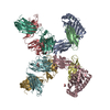 8tq7C  8tq9C 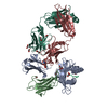 8tqaC  5weuS  8tq4 8tq5 8tq6 C: citing same article ( S: Starting model for refinement |
|---|---|
| Similar structure data |
-Links
 PDBj
PDBj


- Assembly
| Deposited unit | 
| ||||||||||||
|---|---|---|---|---|---|---|---|---|---|---|---|---|---|
| 1 |
| ||||||||||||
| 2 |
| ||||||||||||
| Unit cell |
|
-Components
-Protein , 2 types, 4 molecules ACBD
| #1: Protein | Mass: 31819.361 Da / Num. of mol.: 2 Source method: isolated from a genetically manipulated source Source: (gene. exp.) Mus musculus (house mouse) / Gene: H2-D1 / Plasmid: pET21b / Production host:  Escherichia coli BL21(DE3) (bacteria) / References: UniProt: P01900 Escherichia coli BL21(DE3) (bacteria) / References: UniProt: P01900#2: Protein | Beta-2 microglobulinMass: 11704.359 Da / Num. of mol.: 2 / Mutation: A85D Source method: isolated from a genetically manipulated source Source: (gene. exp.) Mus musculus (house mouse) / Strain: C57BL/6 / Gene: B2m / Plasmid: pET3a / Production host: Escherichia coli BL21(DE3) (bacteria) / References: UniProt: P01887 |
|---|
-Protein/peptide , 1 types, 2 molecules PE
| #3: Protein/peptide | Transmembrane protein / TM / Glycoprotein 41 / gp41 Mass: 1075.265 Da / Num. of mol.: 2 / Fragment: HV1: HIV-1 P18-I10 (UNP residues 311-320) / Source method: obtained synthetically / Source: (synth.) Human immunodeficiency virus 1 / References: UniProt: P04578 |
|---|
-Antibody , 2 types, 4 molecules HFLG
| #4: Antibody | Mass: 23325.117 Da / Num. of mol.: 2 Source method: isolated from a genetically manipulated source Source: (gene. exp.) Mus musculus (house mouse) / Cell line (production host): 34.5.8 / Production host: Mus musculus (house mouse)#5: Antibody | Mass: 24207.707 Da / Num. of mol.: 2 Source method: isolated from a genetically manipulated source Source: (gene. exp.) Mus musculus (house mouse) / Cell line (production host): 34.5.8 / Production host: Mus musculus (house mouse) |
|---|
-Non-polymers , 3 types, 131 molecules 


| #6: Chemical | ChemComp-GOL / Glycerol Mass: 92.094 Da / Num. of mol.: 5 / Source method: obtained synthetically / Formula: C3H8O3 Mass: 92.094 Da / Num. of mol.: 5 / Source method: obtained synthetically / Formula: C3H8O3#7: Chemical | Ethylene glycol Mass: 62.068 Da / Num. of mol.: 3 / Source method: isolated from a natural source / Formula: C2H6O2 Mass: 62.068 Da / Num. of mol.: 3 / Source method: isolated from a natural source / Formula: C2H6O2#8: Water | ChemComp-HOH / | WaterMass: 18.015 Da / Num. of mol.: 123 / Source method: isolated from a natural source / Formula: H2O |
|---|
-Details
| Has ligand of interest | N |
|---|
-Experimental details
-Experiment
| Experiment | Method: X-RAY DIFFRACTION / Number of used crystals: 1 |
|---|
- Sample preparation
| Crystal | Density Matthews: 2.71 Å3/Da / Density % sol: 54.53 % |
|---|---|
| Crystal grow | Temperature: 291 K / Method: vapor diffusion, hanging drop / pH: 6 Details: 18% PEG4000, 0.1 M MES, pH 6.0, 0.12 M calcium acetate |
-Data collection
| Diffraction | Mean temperature: 273 K / Serial crystal experiment: N |
|---|---|
| Diffraction source | Source: SYNCHROTRON / Site: APS / Beamline: 22-ID / Wavelength: 1 Å |
| Detector | Type: DECTRIS EIGER X 16M / Detector: PIXEL / Date: Sep 23, 2022 |
| Radiation | Protocol: SINGLE WAVELENGTH / Monochromatic (M) / Laue (L): M / Scattering type: x-ray |
| Radiation wavelength | Wavelength: 1 Å / Relative weight: 1 |
| Reflection | Resolution: 2.69→87.09 Å / Num. obs: 54120 / % possible obs: 97.9 % / Redundancy: 3.8 % / Biso Wilson estimate: 57.76 Å2 / CC1/2: 0.99 / CC star: 0.997 / Rmerge(I) obs: 0.145 / Rpim(I) all: 0.085 / Net I/σ(I): 7.6 |
| Reflection shell | Resolution: 2.69→2.8 Å / Rmerge(I) obs: 0.927 / Mean I/σ(I) obs: 1.3 / Num. unique obs: 5458 / CC1/2: 0.565 / CC star: 0.85 / Rpim(I) all: 0.455 / % possible all: 99.3 |
- Processing
| Software |
| ||||||||||||||||||||||||||||||||||||||||||||||||||||||||||||||||||||||||||||||||||||||||||||||||||||||||||||||||||||||||||||||||||||||||||||
|---|---|---|---|---|---|---|---|---|---|---|---|---|---|---|---|---|---|---|---|---|---|---|---|---|---|---|---|---|---|---|---|---|---|---|---|---|---|---|---|---|---|---|---|---|---|---|---|---|---|---|---|---|---|---|---|---|---|---|---|---|---|---|---|---|---|---|---|---|---|---|---|---|---|---|---|---|---|---|---|---|---|---|---|---|---|---|---|---|---|---|---|---|---|---|---|---|---|---|---|---|---|---|---|---|---|---|---|---|---|---|---|---|---|---|---|---|---|---|---|---|---|---|---|---|---|---|---|---|---|---|---|---|---|---|---|---|---|---|---|---|---|
| Refinement | Method to determine structure: MOLECULAR REPLACEMENT Starting model: PDB entry 5WEU Resolution: 2.69→87.09 Å / Cross valid method: FREE R-VALUE / σ(F): 154.07 / Phase error: 23.426 Stereochemistry target values: GeoStd + Monomer Library + CDL v1.2
| ||||||||||||||||||||||||||||||||||||||||||||||||||||||||||||||||||||||||||||||||||||||||||||||||||||||||||||||||||||||||||||||||||||||||||||
| Solvent computation | Shrinkage radii: 0.9 Å / VDW probe radii: 1.1 Å / Solvent model: FLAT BULK SOLVENT MODEL | ||||||||||||||||||||||||||||||||||||||||||||||||||||||||||||||||||||||||||||||||||||||||||||||||||||||||||||||||||||||||||||||||||||||||||||
| Displacement parameters | Biso mean: 60.4 Å2 | ||||||||||||||||||||||||||||||||||||||||||||||||||||||||||||||||||||||||||||||||||||||||||||||||||||||||||||||||||||||||||||||||||||||||||||
| Refinement step | Cycle: LAST / Resolution: 2.69→87.09 Å
| ||||||||||||||||||||||||||||||||||||||||||||||||||||||||||||||||||||||||||||||||||||||||||||||||||||||||||||||||||||||||||||||||||||||||||||
| Refine LS restraints |
| ||||||||||||||||||||||||||||||||||||||||||||||||||||||||||||||||||||||||||||||||||||||||||||||||||||||||||||||||||||||||||||||||||||||||||||
| LS refinement shell |
|
