 Movie
Movie Controller

[English] 日本語
 Yorodumi
- PDB-8tkn: Murine NF-kappaB p50 Rel Homology Region homodimer in complex wit... -
+ Open data

- Basic information
| Entry | Database: PDB / ID: 8tkn | ||||||
|---|---|---|---|---|---|---|---|
| Title | Murine NF-kappaB p50 Rel Homology Region homodimer in complex with 10-mer kappaB DNA from human Neutrophil Gelatinase-associated Lipocalin (NGAL) promoter | ||||||
 Components Components |
| ||||||
 Keywords Keywords | DNA BINDING PROTEIN/DNA /  DNA / NF-kappaB / p50 / DNA BINDING PROTEIN / DNA BINDING PROTEIN-DNA complex DNA / NF-kappaB / p50 / DNA BINDING PROTEIN / DNA BINDING PROTEIN-DNA complex | ||||||
| Function / homology |  Function and homology information Function and homology informationcellular response to diterpene / cellular response to glucoside / Regulated proteolysis of p75NTR / Interleukin-1 processing / DEx/H-box helicases activate type I IFN and inflammatory cytokines production / RIP-mediated NFkB activation via ZBP1 / MAP3K8 (TPL2)-dependent MAPK1/3 activation / TRAF6 mediated NF-kB activation / positive regulation of hyaluronan biosynthetic process / NF-kB is activated and signals survival ...cellular response to diterpene / cellular response to glucoside / Regulated proteolysis of p75NTR / Interleukin-1 processing / DEx/H-box helicases activate type I IFN and inflammatory cytokines production / RIP-mediated NFkB activation via ZBP1 / MAP3K8 (TPL2)-dependent MAPK1/3 activation / TRAF6 mediated NF-kB activation / positive regulation of hyaluronan biosynthetic process / NF-kB is activated and signals survival / antibacterial innate immune response / PKMTs methylate histone lysines / Activation of NF-kappaB in B cells / TAK1-dependent IKK and NF-kappa-B activation / cellular response to carbohydrate stimulus / mammary gland involution / FCERI mediated NF-kB activation / CLEC7A (Dectin-1) signaling / Interleukin-1 signaling / cellular response to peptide / cellular response to interleukin-17 / Downstream TCR signaling / NF-kappaB p50/p65 complex / CD209 (DC-SIGN) signaling / negative regulation of interleukin-12 production / cellular response to dsRNA / cellular response to interleukin-6 / actinin binding / cellular response to angiotensin / negative regulation of cytokine production / positive regulation of miRNA metabolic process / non-canonical NF-kappaB signal transduction / cellular response to cytokine stimulus / cellular response to organic cyclic compound / positive regulation of transcription initiation by RNA polymerase II / canonical NF-kappaB signal transduction / lymph node development / cellular response to interleukin-1 / JNK cascade / cellular response to brain-derived neurotrophic factor stimulus / heat shock protein binding / response to muscle stretch / Neutrophil degranulation / protein sequestering activity / response to cytokine / transcription coregulator activity / RNA polymerase II transcription regulatory region sequence-specific DNA binding / B cell receptor signaling pathway / response to organic cyclic compound / cellular response to virus / DNA-binding transcription repressor activity, RNA polymerase II-specific / negative regulation of inflammatory response / cellular response to mechanical stimulus / cellular response to nicotine / positive regulation of canonical Wnt signaling pathway / MAPK cascade / sequence-specific double-stranded DNA binding / cellular response to tumor necrosis factor / gene expression / double-stranded DNA binding / DNA-binding transcription activator activity, RNA polymerase II-specific / response to oxidative stress / cellular response to lipopolysaccharide / transcription regulator complex / sequence-specific DNA binding / transcription cis-regulatory region binding / DNA-binding transcription factor activity, RNA polymerase II-specific / inflammatory response / RNA polymerase II cis-regulatory region sequence-specific DNA binding / DNA-binding transcription factor activity / negative regulation of gene expression / innate immune response / negative regulation of DNA-templated transcription / apoptotic process / chromatin binding / chromatin / protein-containing complex binding / positive regulation of gene expression / regulation of transcription by RNA polymerase II / negative regulation of apoptotic process / positive regulation of DNA-templated transcription / negative regulation of transcription by RNA polymerase II / positive regulation of transcription by RNA polymerase II / protein-containing complex / mitochondrion / DNA binding / nucleoplasm / identical protein binding / nucleus / cytosol / cytoplasmSimilarity search - Function | ||||||
| Biological species |  Mus musculus (house mouse) Mus musculus (house mouse) Homo sapiens (human) Homo sapiens (human) | ||||||
| Method | X-RAY DIFFRACTION / SYNCHROTRON / MOLECULAR REPLACEMENT / Resolution: 2.8 Å | ||||||
 Authors Authors | Zhu, N. / Mealka, M. / Mitchel, S. / Rogers, W.E. / Huxford, T. | ||||||
| Funding support | 1items
| ||||||
 Citation Citation | Journal: Biomolecules / Year: 2023 Title: X-ray Crystallographic Study of Preferred Spacing by the NF-kappa B p50 Homodimer on kappa B DNA. Authors: Zhu, N. / Mealka, M. / Mitchel, S. / Milani, C. / Acuna, L.M. / Rogers, E. / Lahana, A.N. / Huxford, T. | ||||||
| History |
|
- Structure visualization
| Structure viewer | Molecule: MolmilJmol/JSmol |
|---|
- Downloads & links
-Download
| PDBx/mmCIF format | 8tkn.cif.gz | 190.2 KB | Display | PDBx/mmCIF format |
|---|---|---|---|---|
| PDB format | pdb8tkn.ent.gz | 119.8 KB | Display | PDB format |
| PDBx/mmJSON format | 8tkn.json.gz | Tree view | PDBx/mmJSON format | |
| Others |  Other downloads Other downloads |
-Validation report
| Arichive directory | https://data.pdbj.org/pub/pdb/validation_reports/tk/8tknftp://data.pdbj.org/pub/pdb/validation_reports/tk/8tkn | HTTPS FTP |
|---|
-Related structure data
| Related structure data | 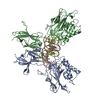 8tklC 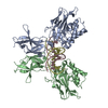 8tkmC 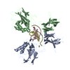 1nfkS S: Starting model for refinement C: citing same article ( |
|---|---|
| Similar structure data |
-Links
 PDBj
PDBj


- Assembly
| Deposited unit | 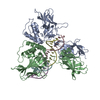
| ||||||||||||
|---|---|---|---|---|---|---|---|---|---|---|---|---|---|
| 1 |
| ||||||||||||
| Unit cell |
|
-Components
| #1: Protein | Mass: 34924.871 Da / Num. of mol.: 2 Source method: isolated from a genetically manipulated source Source: (gene. exp.) Mus musculus (house mouse) / Gene: Nfkb1 / Production host:  Escherichia coli BL21(DE3) (bacteria) / References: UniProt: P25799 Escherichia coli BL21(DE3) (bacteria) / References: UniProt: P25799#2: DNA chain | Mass: 3085.029 Da / Num. of mol.: 2 / Source method: obtained synthetically / Source: (synth.) Homo sapiens (human)#3: DNA chain | Mass: 3004.981 Da / Num. of mol.: 2 / Source method: obtained synthetically / Source: (synth.) Homo sapiens (human) |
|---|
-Experimental details
-Experiment
| Experiment | Method: X-RAY DIFFRACTION / Number of used crystals: 1 |
|---|
- Sample preparation
| Crystal | Density Matthews: 2.68 Å3/Da / Density % sol: 54.05 % |
|---|---|
| Crystal grow | Temperature: 298 K / Method: vapor diffusion, hanging drop / pH: 5.4 Details: 0.05 M sodium acetate pH 5.4, 20 mM magnesium sulfate, and 4% PEG 3350 Temp details: Room temperature |
-Data collection
| Diffraction | Mean temperature: 100 K / Serial crystal experiment: N |
|---|---|
| Diffraction source | Source: SYNCHROTRON / Site: ALS  / Beamline: 8.2.1 / Wavelength: 0.99999 Å / Beamline: 8.2.1 / Wavelength: 0.99999 Å |
| Detector | Type: ADSC QUANTUM 315r / Detector: CCD / Date: Oct 22, 2011 |
| Radiation | Protocol: SINGLE WAVELENGTH / Monochromatic (M) / Laue (L): M / Scattering type: x-ray |
| Radiation wavelength | Wavelength: 0.99999 Å / Relative weight: 1 |
| Reflection | Resolution: 2.8→50 Å / Num. obs: 23123 / % possible obs: 100 % / Redundancy: 6 % / Biso Wilson estimate: 74.81 Å2 / Rsym value: 0.083 / Net I/σ(I): 19 |
| Reflection shell | Resolution: 2.8→2.85 Å / Mean I/σ(I) obs: 2.3 / Num. unique obs: 2310 / Rsym value: 0.764 |
- Processing
| Software |
| |||||||||||||||||||||||||||||||||||||||||||||||||||||||||||||||
|---|---|---|---|---|---|---|---|---|---|---|---|---|---|---|---|---|---|---|---|---|---|---|---|---|---|---|---|---|---|---|---|---|---|---|---|---|---|---|---|---|---|---|---|---|---|---|---|---|---|---|---|---|---|---|---|---|---|---|---|---|---|---|---|---|
| Refinement | Method to determine structure: MOLECULAR REPLACEMENT Starting model: 1NFK Resolution: 2.8→47.62 Å / SU ML: 0.4335 / Cross valid method: FREE R-VALUE / σ(F): 1.34 / Phase error: 32.0864 Stereochemistry target values: GeoStd + Monomer Library + CDL v1.2
| |||||||||||||||||||||||||||||||||||||||||||||||||||||||||||||||
| Solvent computation | Shrinkage radii: 0.9 Å / VDW probe radii: 1.1 Å / Solvent model: FLAT BULK SOLVENT MODEL | |||||||||||||||||||||||||||||||||||||||||||||||||||||||||||||||
| Displacement parameters | Biso mean: 81.31 Å2 | |||||||||||||||||||||||||||||||||||||||||||||||||||||||||||||||
| Refinement step | Cycle: LAST / Resolution: 2.8→47.62 Å
| |||||||||||||||||||||||||||||||||||||||||||||||||||||||||||||||
| Refine LS restraints |
| |||||||||||||||||||||||||||||||||||||||||||||||||||||||||||||||
| LS refinement shell |
|
