 Movie
Movie Controller

[English] 日本語
 Yorodumi
- PDB-8thm: Beta carbonic anhydrase from the carboxysome of Cyanobium PCC 7001 -
+ Open data

- Basic information
| Entry | Database: PDB / ID: 8thm | ||||||
|---|---|---|---|---|---|---|---|
| Title | Beta carbonic anhydrase from the carboxysome of Cyanobium PCC 7001 | ||||||
 Components Components | Carboxysome shell carbonic anhydrase | ||||||
 Keywords Keywords |  PHOTOSYNTHESIS / beta-carbonic anhydrase / carboxysome / cyanobacteria / RuBP activated / LYASE PHOTOSYNTHESIS / beta-carbonic anhydrase / carboxysome / cyanobacteria / RuBP activated / LYASE | ||||||
| Function / homology |  Function and homology informationcarboxysome / carbon fixation / carbonic anhydrase / carbonate dehydratase activity / metal ion binding Function and homology informationcarboxysome / carbon fixation / carbonic anhydrase / carbonate dehydratase activity / metal ion bindingSimilarity search - Function | ||||||
| Biological species |  Cyanobium sp. PCC 7001 (bacteria) Cyanobium sp. PCC 7001 (bacteria) | ||||||
| Method | X-RAY DIFFRACTION / SYNCHROTRON / MOLECULAR REPLACEMENT / Resolution: 2.3 Å | ||||||
 Authors Authors | Pulsford, S.B. / Jackson, C.J. | ||||||
| Funding support |  Australia, 1items Australia, 1items
| ||||||
 Citation Citation | Journal: To Be Published Title: Alpha-cyanobacterial carbonic anhydrase is allosterically regulated by the Rubisco substrate Ribulose 1,5-bisphosphate (RuBP) Authors: Pulsford, S.B. / Outram, M.A. / Forster, B. / Jackson, C.J. / Rhodes, T. / Williams, S.J. / Price, G.D. / Badger, M.R. / Long, B.M. | ||||||
| History |
|
- Structure visualization
| Structure viewer | Molecule: MolmilJmol/JSmol |
|---|
- Downloads & links
-Download
| PDBx/mmCIF format | 8thm.cif.gz | 562.8 KB | Display | PDBx/mmCIF format |
|---|---|---|---|---|
| PDB format | pdb8thm.ent.gz | 464.7 KB | Display | PDB format |
| PDBx/mmJSON format | 8thm.json.gz | Tree view | PDBx/mmJSON format | |
| Others |  Other downloads Other downloads |
-Validation report
| Arichive directory | https://data.pdbj.org/pub/pdb/validation_reports/th/8thmftp://data.pdbj.org/pub/pdb/validation_reports/th/8thm | HTTPS FTP |
|---|
-Related structure data
| Similar structure data |
|---|
-Links
 PDBj
PDBj


- Assembly
| Deposited unit | 
| ||||||||
|---|---|---|---|---|---|---|---|---|---|
| 1 |
| ||||||||
| Unit cell |
|
-Components
-Protein / Sugars , 2 types, 12 molecules ABCDEF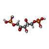
| #1: Protein | Mass: 51308.566 Da / Num. of mol.: 6 Source method: isolated from a genetically manipulated source Source: (gene. exp.) Cyanobium sp. PCC 7001 (bacteria) / Gene: CPCC7001_23 / Production host: Escherichia coli BL21(DE3) (bacteria) / References: UniProt: B5ILN4, carbonic anhydrase#6: Sugar | ChemComp-RUB / Ribulose 1,5-bisphosphate Type: saccharideCarbohydrate / Mass: 310.090 Da / Num. of mol.: 6 / Source method: obtained synthetically / Formula: C5H12O11P2 / Feature type: SUBJECT OF INVESTIGATION Type: saccharideCarbohydrate / Mass: 310.090 Da / Num. of mol.: 6 / Source method: obtained synthetically / Formula: C5H12O11P2 / Feature type: SUBJECT OF INVESTIGATION |
|---|
-Non-polymers , 7 types, 815 molecules 


| #2: Chemical | ChemComp-ZN /  Mass: 65.409 Da / Num. of mol.: 8 / Source method: obtained synthetically / Formula: Zn / Feature type: SUBJECT OF INVESTIGATION Mass: 65.409 Da / Num. of mol.: 8 / Source method: obtained synthetically / Formula: Zn / Feature type: SUBJECT OF INVESTIGATION#3: Chemical | Glycerol Mass: 92.094 Da / Num. of mol.: 3 / Source method: obtained synthetically / Formula: C3H8O3 Mass: 92.094 Da / Num. of mol.: 3 / Source method: obtained synthetically / Formula: C3H8O3#4: Chemical | ChemComp-SO4 / Sulfate Mass: 96.063 Da / Num. of mol.: 18 / Source method: obtained synthetically / Formula: SO4 Mass: 96.063 Da / Num. of mol.: 18 / Source method: obtained synthetically / Formula: SO4#5: Chemical | ChemComp-EDO / Ethylene glycol Mass: 62.068 Da / Num. of mol.: 10 / Source method: obtained synthetically / Formula: C2H6O2 Mass: 62.068 Da / Num. of mol.: 10 / Source method: obtained synthetically / Formula: C2H6O2#7: Chemical | Carbon dioxide Mass: 44.010 Da / Num. of mol.: 2 / Source method: obtained synthetically / Formula: CO2 Mass: 44.010 Da / Num. of mol.: 2 / Source method: obtained synthetically / Formula: CO2#8: Chemical | ChemComp-BCT / | Bicarbonate Mass: 61.017 Da / Num. of mol.: 1 / Source method: isolated from a natural source / Formula: CHO3 / Comment: pH buffer*YM Mass: 61.017 Da / Num. of mol.: 1 / Source method: isolated from a natural source / Formula: CHO3 / Comment: pH buffer*YM#9: Water | ChemComp-HOH / | WaterMass: 18.015 Da / Num. of mol.: 773 / Source method: isolated from a natural source / Formula: H2O |
|---|
-Details
| Has ligand of interest | Y |
|---|
-Experimental details
-Experiment
| Experiment | Method: X-RAY DIFFRACTION / Number of used crystals: 1 |
|---|
- Sample preparation
| Crystal | Density Matthews: 2.93 Å3/Da / Density % sol: 58.03 % |
|---|---|
| Crystal grow | Temperature: 293.15 K / Method: vapor diffusion, hanging drop / pH: 6.5 Details: 0.2M Ammonium sulfate, 0.1M BIS-TRIS (pH 6.5), 21% PEG 3350, 15% Ethylene glycol |
-Data collection
| Diffraction | Mean temperature: 100 K / Serial crystal experiment: N |
|---|---|
| Diffraction source | Source: SYNCHROTRON / Site: Australian Synchrotron / Beamline: MX2 / Wavelength: 0.9537 Å |
| Detector | Type: DECTRIS EIGER X 16M / Detector: PIXEL / Date: Oct 29, 2021 |
| Radiation | Protocol: SINGLE WAVELENGTH / Monochromatic (M) / Laue (L): M / Scattering type: x-ray |
| Radiation wavelength | Wavelength: 0.9537 Å / Relative weight: 1 |
| Reflection | Resolution: 2.3→48.48 Å / Num. obs: 160617 / % possible obs: 100 % / Redundancy: 12.3 % / CC1/2: 0.997 / Rmerge(I) obs: 0.26 / Rpim(I) all: 0.077 / Rrim(I) all: 0.271 / Χ2: 1.02 / Net I/σ(I): 8.1 / Num. measured all: 1970240 |
| Reflection shell | Resolution: 2.3→2.34 Å / % possible obs: 100 % / Redundancy: 12.7 % / Rmerge(I) obs: 5.093 / Num. measured all: 100213 / Num. unique obs: 7868 / CC1/2: 0.297 / Rpim(I) all: 1.468 / Rrim(I) all: 5.302 / Χ2: 1.04 / Net I/σ(I) obs: 0.7 |
- Processing
| Software |
| |||||||||||||||||||||||||||||||||||||||||||||||||||||||||||||||||||||||||||||||||||||||||||||||||||||||||||||||||||||||||||||||||||||||||||||||||||||||||||||||||||||||||||||||||||||||||||||||||||||||||||||||||||||||||
|---|---|---|---|---|---|---|---|---|---|---|---|---|---|---|---|---|---|---|---|---|---|---|---|---|---|---|---|---|---|---|---|---|---|---|---|---|---|---|---|---|---|---|---|---|---|---|---|---|---|---|---|---|---|---|---|---|---|---|---|---|---|---|---|---|---|---|---|---|---|---|---|---|---|---|---|---|---|---|---|---|---|---|---|---|---|---|---|---|---|---|---|---|---|---|---|---|---|---|---|---|---|---|---|---|---|---|---|---|---|---|---|---|---|---|---|---|---|---|---|---|---|---|---|---|---|---|---|---|---|---|---|---|---|---|---|---|---|---|---|---|---|---|---|---|---|---|---|---|---|---|---|---|---|---|---|---|---|---|---|---|---|---|---|---|---|---|---|---|---|---|---|---|---|---|---|---|---|---|---|---|---|---|---|---|---|---|---|---|---|---|---|---|---|---|---|---|---|---|---|---|---|---|---|---|---|---|---|---|---|---|---|---|---|---|---|---|---|---|
| Refinement | Method to determine structure: MOLECULAR REPLACEMENT / Resolution: 2.3→47.51 Å / SU ML: 0.38 / Cross valid method: FREE R-VALUE / σ(F): 1.33 / Phase error: 29.48 / Stereochemistry target values: ML
| |||||||||||||||||||||||||||||||||||||||||||||||||||||||||||||||||||||||||||||||||||||||||||||||||||||||||||||||||||||||||||||||||||||||||||||||||||||||||||||||||||||||||||||||||||||||||||||||||||||||||||||||||||||||||
| Solvent computation | Shrinkage radii: 0.9 Å / VDW probe radii: 1.11 Å / Solvent model: FLAT BULK SOLVENT MODEL | |||||||||||||||||||||||||||||||||||||||||||||||||||||||||||||||||||||||||||||||||||||||||||||||||||||||||||||||||||||||||||||||||||||||||||||||||||||||||||||||||||||||||||||||||||||||||||||||||||||||||||||||||||||||||
| Refinement step | Cycle: LAST / Resolution: 2.3→47.51 Å
| |||||||||||||||||||||||||||||||||||||||||||||||||||||||||||||||||||||||||||||||||||||||||||||||||||||||||||||||||||||||||||||||||||||||||||||||||||||||||||||||||||||||||||||||||||||||||||||||||||||||||||||||||||||||||
| Refine LS restraints |
| |||||||||||||||||||||||||||||||||||||||||||||||||||||||||||||||||||||||||||||||||||||||||||||||||||||||||||||||||||||||||||||||||||||||||||||||||||||||||||||||||||||||||||||||||||||||||||||||||||||||||||||||||||||||||
| LS refinement shell |
|
