 Movie
Movie Controller

[English] 日本語
 Yorodumi
- PDB-8tcu: Structure of PYCR1 complexed with 2-chloro-5-(2-oxoimidazolidin-1... -
+ Open data

- Basic information
| Entry | Database: PDB / ID: 8tcu | ||||||
|---|---|---|---|---|---|---|---|
| Title | Structure of PYCR1 complexed with 2-chloro-5-(2-oxoimidazolidin-1-yl)benzoic acid | ||||||
 Components Components | Pyrroline-5-carboxylate reductase 1, mitochondrial | ||||||
 Keywords Keywords | OXIDOREDUCTASE / AMINO-ACID BIOSYNTHESIS / PROLINE BIOSYNTHESIS | ||||||
| Function / homology |  Function and homology informationpyrroline-5-carboxylate reductase / pyrroline-5-carboxylate reductase activity / L-proline biosynthetic process / Glutamate and glutamine metabolism / proline biosynthetic process / negative regulation of oxidative stress-induced neuron intrinsic apoptotic signaling pathway / regulation of mitochondrial membrane potential / cellular response to oxidative stress / mitochondrial matrix / mitochondrion / identical protein binding Function and homology informationpyrroline-5-carboxylate reductase / pyrroline-5-carboxylate reductase activity / L-proline biosynthetic process / Glutamate and glutamine metabolism / proline biosynthetic process / negative regulation of oxidative stress-induced neuron intrinsic apoptotic signaling pathway / regulation of mitochondrial membrane potential / cellular response to oxidative stress / mitochondrial matrix / mitochondrion / identical protein bindingSimilarity search - Function | ||||||
| Biological species |  Homo sapiens (human) Homo sapiens (human) | ||||||
| Method | X-RAY DIFFRACTION / SYNCHROTRON / FOURIER SYNTHESIS / Resolution: 2 Å | ||||||
 Authors Authors | Tanner, J.J. / Meeks, K.R. | ||||||
| Funding support |  United States, 1items United States, 1items
| ||||||
 Citation Citation | Journal: J.Chem.Inf.Model. / Year: 2024 Title: Novel Fragment Inhibitors of PYCR1 from Docking-Guided X-ray Crystallography. Authors: Meeks, K.R. / Ji, J. / Protopopov, M.V. / Tarkhanova, O.O. / Moroz, Y.S. / Tanner, J.J. | ||||||
| History |
|
- Structure visualization
| Structure viewer | Molecule: MolmilJmol/JSmol |
|---|
- Downloads & links
-Download
| PDBx/mmCIF format | 8tcu.cif.gz | 509 KB | Display | PDBx/mmCIF format |
|---|---|---|---|---|
| PDB format | pdb8tcu.ent.gz | 418 KB | Display | PDB format |
| PDBx/mmJSON format | 8tcu.json.gz | Tree view | PDBx/mmJSON format | |
| Others |  Other downloads Other downloads |
-Validation report
| Arichive directory | https://data.pdbj.org/pub/pdb/validation_reports/tc/8tcuftp://data.pdbj.org/pub/pdb/validation_reports/tc/8tcu | HTTPS FTP |
|---|
-Related structure data
| Related structure data |  8tcvC 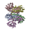 8tcwC  8tcxC  8tcyC 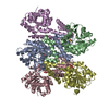 8tczC 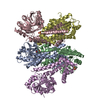 8td0C  8td1C C: citing same article ( |
|---|---|
| Similar structure data |
-Links
 PDBj
PDBj


- Assembly
| Deposited unit | 
| ||||||||||||
|---|---|---|---|---|---|---|---|---|---|---|---|---|---|
| 1 | 
| ||||||||||||
| Unit cell |
| ||||||||||||
| Components on special symmetry positions |
|
-Components
| #1: Protein | / P5C reductase 1 / P5CR 1 Mass: 33532.574 Da / Num. of mol.: 5 Source method: isolated from a genetically manipulated source Source: (gene. exp.) Homo sapiens (human) / Gene: PYCR1 / Production host:  Escherichia coli (E. coli) Escherichia coli (E. coli)References: UniProt: P32322, pyrroline-5-carboxylate reductase#2: Chemical | ChemComp-ZR3 / Mass: 240.643 Da / Num. of mol.: 5 / Source method: obtained synthetically / Formula: C10H9ClN2O3 / Feature type: SUBJECT OF INVESTIGATION #3: Chemical | ChemComp-SO4 / Sulfate  Mass: 96.063 Da / Num. of mol.: 5 / Source method: obtained synthetically / Formula: SO4 Mass: 96.063 Da / Num. of mol.: 5 / Source method: obtained synthetically / Formula: SO4#4: Water | ChemComp-HOH / | Water Mass: 18.015 Da / Num. of mol.: 397 / Source method: isolated from a natural source / Formula: H2O Mass: 18.015 Da / Num. of mol.: 397 / Source method: isolated from a natural source / Formula: H2OHas ligand of interest | Y | |
|---|
-Experimental details
-Experiment
| Experiment | Method: X-RAY DIFFRACTION / Number of used crystals: 1 |
|---|
- Sample preparation
| Crystal | Density Matthews: 2.41 Å3/Da / Density % sol: 48.99 % |
|---|---|
| Crystal grow | Temperature: 293 K / Method: vapor diffusion, sitting drop Details: Reservoir contained 350 mM Li2SO4, 20% (w/v) PEG 3350, and 0.1 M HEPES at pH 7.5. Enzyme solution contained 16 mM 2-chloro-5-(2-oxoimidazolidin-1-yl)benzoic acid. Crystal was soaked in ...Details: Reservoir contained 350 mM Li2SO4, 20% (w/v) PEG 3350, and 0.1 M HEPES at pH 7.5. Enzyme solution contained 16 mM 2-chloro-5-(2-oxoimidazolidin-1-yl)benzoic acid. Crystal was soaked in cryobuffer containing 0 mM Li2SO4, 20% PEG 200, and 25 mM 2-chloro-5-(2-oxoimidazolidin-1-yl)benzoic acid |
-Data collection
| Diffraction | Mean temperature: 100 K / Serial crystal experiment: N |
|---|---|
| Diffraction source | Source: SYNCHROTRON / Site: APS / Beamline: 24-ID-C / Wavelength: 0.97918 Å |
| Detector | Type: DECTRIS EIGER X 16M / Detector: PIXEL / Date: Feb 18, 2023 |
| Radiation | Protocol: SINGLE WAVELENGTH / Monochromatic (M) / Laue (L): M / Scattering type: x-ray |
| Radiation wavelength | Wavelength: 0.97918 Å / Relative weight: 1 |
| Reflection | Resolution: 2→89.46 Å / Num. obs: 103502 / % possible obs: 95.6 % / Redundancy: 2.2 % / CC1/2: 0.983 / Rmerge(I) obs: 0.127 / Rpim(I) all: 0.106 / Rrim(I) all: 0.166 / Χ2: 1.34 / Net I/σ(I): 9.4 / Num. measured all: 232533 |
| Reflection shell | Resolution: 2→2.03 Å / % possible obs: 95.2 % / Redundancy: 2.2 % / Rmerge(I) obs: 0.768 / Num. measured all: 11044 / Num. unique obs: 5121 / CC1/2: 0.412 / Rpim(I) all: 0.638 / Rrim(I) all: 1.003 / Χ2: 0.99 / Net I/σ(I) obs: 1.3 |
- Processing
| Software |
| |||||||||||||||||||||||||||||||||||||||||||||||||||||||||||||||||||||||||||||||||||||||||||||||||||||||||||||||||||||||||||||||||||||||||||||||||||||||||||||||||||||||||||||||||||||||||||||||||||||||||||||||||||||||||
|---|---|---|---|---|---|---|---|---|---|---|---|---|---|---|---|---|---|---|---|---|---|---|---|---|---|---|---|---|---|---|---|---|---|---|---|---|---|---|---|---|---|---|---|---|---|---|---|---|---|---|---|---|---|---|---|---|---|---|---|---|---|---|---|---|---|---|---|---|---|---|---|---|---|---|---|---|---|---|---|---|---|---|---|---|---|---|---|---|---|---|---|---|---|---|---|---|---|---|---|---|---|---|---|---|---|---|---|---|---|---|---|---|---|---|---|---|---|---|---|---|---|---|---|---|---|---|---|---|---|---|---|---|---|---|---|---|---|---|---|---|---|---|---|---|---|---|---|---|---|---|---|---|---|---|---|---|---|---|---|---|---|---|---|---|---|---|---|---|---|---|---|---|---|---|---|---|---|---|---|---|---|---|---|---|---|---|---|---|---|---|---|---|---|---|---|---|---|---|---|---|---|---|---|---|---|---|---|---|---|---|---|---|---|---|---|---|---|---|
| Refinement | Method to determine structure: FOURIER SYNTHESIS / Resolution: 2→89.46 Å / SU ML: 0.24 / Cross valid method: FREE R-VALUE / σ(F): 1.37 / Phase error: 23.29 / Stereochemistry target values: ML
| |||||||||||||||||||||||||||||||||||||||||||||||||||||||||||||||||||||||||||||||||||||||||||||||||||||||||||||||||||||||||||||||||||||||||||||||||||||||||||||||||||||||||||||||||||||||||||||||||||||||||||||||||||||||||
| Solvent computation | Shrinkage radii: 0.9 Å / VDW probe radii: 1.1 Å / Solvent model: FLAT BULK SOLVENT MODEL | |||||||||||||||||||||||||||||||||||||||||||||||||||||||||||||||||||||||||||||||||||||||||||||||||||||||||||||||||||||||||||||||||||||||||||||||||||||||||||||||||||||||||||||||||||||||||||||||||||||||||||||||||||||||||
| Refinement step | Cycle: LAST / Resolution: 2→89.46 Å
| |||||||||||||||||||||||||||||||||||||||||||||||||||||||||||||||||||||||||||||||||||||||||||||||||||||||||||||||||||||||||||||||||||||||||||||||||||||||||||||||||||||||||||||||||||||||||||||||||||||||||||||||||||||||||
| Refine LS restraints |
| |||||||||||||||||||||||||||||||||||||||||||||||||||||||||||||||||||||||||||||||||||||||||||||||||||||||||||||||||||||||||||||||||||||||||||||||||||||||||||||||||||||||||||||||||||||||||||||||||||||||||||||||||||||||||
| LS refinement shell |
| |||||||||||||||||||||||||||||||||||||||||||||||||||||||||||||||||||||||||||||||||||||||||||||||||||||||||||||||||||||||||||||||||||||||||||||||||||||||||||||||||||||||||||||||||||||||||||||||||||||||||||||||||||||||||
| Refinement TLS params. | Method: refined / Refine-ID: X-RAY DIFFRACTION
| |||||||||||||||||||||||||||||||||||||||||||||||||||||||||||||||||||||||||||||||||||||||||||||||||||||||||||||||||||||||||||||||||||||||||||||||||||||||||||||||||||||||||||||||||||||||||||||||||||||||||||||||||||||||||
| Refinement TLS group |
|
