 Movie
Movie Controller

[English] 日本語
 Yorodumi
- PDB-8tc1: Cryo-EM Structure of Spike Glycoprotein from Civet Coronavirus 00... -
+ Open data

- Basic information
| Entry | Database: PDB / ID: 8tc1 | ||||||
|---|---|---|---|---|---|---|---|
| Title | Cryo-EM Structure of Spike Glycoprotein from Civet Coronavirus 007 in Closed Conformation | ||||||
 Components Components | Spike glycoprotein Spike protein Spike protein | ||||||
 Keywords Keywords | VIRAL PROTEIN / Spike / Glycoprotein / Coronavirus | ||||||
| Function / homology |  Function and homology information Function and homology informationendocytosis involved in viral entry into host cell / host cell endoplasmic reticulum-Golgi intermediate compartment membrane / receptor-mediated virion attachment to host cell / fusion of virus membrane with host plasma membrane / fusion of virus membrane with host endosome membrane / viral envelope / host cell plasma membrane / virion membrane / membraneSimilarity search - Function | ||||||
| Biological species |  Paguma larvata (masked palm civet) Paguma larvata (masked palm civet) | ||||||
| Method | ELECTRON MICROSCOPY / single particle reconstruction / cryo EM / Resolution: 1.92 Å | ||||||
 Authors Authors | Bostina, M. / Hills, F.R. / Eruera, A.R. | ||||||
| Funding support |  New Zealand, 1items New Zealand, 1items
| ||||||
 Citation Citation | Journal: To Be Published Title: Cryo-EM reveals variationo in structural motifs within animal SARS-related coronavirus spike protein Authors: Bostina, M. / Hills, F.R. / Eruera, A.R. | ||||||
| History |
|
- Structure visualization
| Structure viewer | Molecule: MolmilJmol/JSmol |
|---|
- Downloads & links
-Download
| PDBx/mmCIF format | 8tc1.cif.gz | 1.4 MB | Display | PDBx/mmCIF format |
|---|---|---|---|---|
| PDB format | pdb8tc1.ent.gz | 987.3 KB | Display | PDB format |
| PDBx/mmJSON format | 8tc1.json.gz | Tree view | PDBx/mmJSON format | |
| Others |  Other downloads Other downloads |
-Validation report
| Arichive directory | https://data.pdbj.org/pub/pdb/validation_reports/tc/8tc1ftp://data.pdbj.org/pub/pdb/validation_reports/tc/8tc1 | HTTPS FTP |
|---|
-Related structure data
| Related structure data | 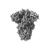 41150MC M: map data used to model this data C: citing same article ( |
|---|---|
| Similar structure data |
-Links
 PDBj
PDBj
- Assembly
| Deposited unit | 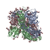
|
|---|---|
| 1 |
|
-Components
-Protein , 1 types, 3 molecules ABC
| #1: Protein | Spike protein / S glycoprotein / E2 / Peplomer protein Mass: 122479.953 Da / Num. of mol.: 3 Source method: isolated from a genetically manipulated source Source: (gene. exp.) Paguma larvata (masked palm civet) / Production host:  Cricetulus griseus (Chinese hamster) / References: UniProt: Q3ZTF3 Cricetulus griseus (Chinese hamster) / References: UniProt: Q3ZTF3 |
|---|
-Sugars , 3 types, 45 molecules 
| #2: Polysaccharide | 2-acetamido-2-deoxy-beta-D-glucopyranose-(1-4)-2-acetamido-2-deoxy-beta-D-glucopyranose / Mass: 424.401 Da / Num. of mol.: 21Source method: isolated from a genetically manipulated source #3: Polysaccharide | beta-D-mannopyranose-(1-4)-2-acetamido-2-deoxy-beta-D-glucopyranose-(1-4)-2-acetamido-2-deoxy-beta- ...beta-D-mannopyranose-(1-4)-2-acetamido-2-deoxy-beta-D-glucopyranose-(1-4)-2-acetamido-2-deoxy-beta-D-glucopyranose / Mass: 586.542 Da / Num. of mol.: 9Source method: isolated from a genetically manipulated source #4: Sugar | ChemComp-NAG / N-Acetylglucosamine Type: D-saccharide, beta linking / Mass: 221.208 Da / Num. of mol.: 15 / Source method: obtained synthetically / Formula: C8H15NO6 Type: D-saccharide, beta linking / Mass: 221.208 Da / Num. of mol.: 15 / Source method: obtained synthetically / Formula: C8H15NO6 |
|---|
-Non-polymers , 2 types, 585 molecules 

| #5: Chemical | Linoleic acid Mass: 280.445 Da / Num. of mol.: 3 / Source method: obtained synthetically / Formula: C18H32O2 Mass: 280.445 Da / Num. of mol.: 3 / Source method: obtained synthetically / Formula: C18H32O2#6: Water | ChemComp-HOH / | WaterMass: 18.015 Da / Num. of mol.: 582 / Source method: isolated from a natural source / Formula: H2O |
|---|
-Details
| Has ligand of interest | N |
|---|
-Experimental details
-Experiment
| Experiment | Method: ELECTRON MICROSCOPY |
|---|---|
| EM experiment | Aggregation state: PARTICLE / 3D reconstruction method: single particle reconstruction |
- Sample preparation
| Component | Name: Civet SARS CoV 007/2004 / Type: VIRUS / Entity ID: #1 / Source: RECOMBINANT |
|---|---|
| Source (natural) | Organism:  Civet SARS CoV 007/2004 (virus) Civet SARS CoV 007/2004 (virus) |
| Source (recombinant) | Organism: Cricetulus griseus (Chinese hamster) |
| Details of virus | Empty: NO / Enveloped: YES / Isolate: STRAIN / Type: VIRION |
| Buffer solution | pH: 8 |
| Specimen | Embedding applied: NO / Shadowing applied: NO / Staining applied: NO / Vitrification applied: YES |
| Vitrification | Cryogen name: ETHANE |
- Electron microscopy imaging
| Experimental equipment |  Model: Titan Krios / Image courtesy: FEI Company |
|---|---|
| Microscopy | Model: FEI TITAN KRIOS |
| Electron gun | Electron source: FIELD EMISSION GUN / Accelerating voltage: 300 kV / Illumination mode: SPOT SCAN |
| Electron lens | Mode: OTHER / Nominal defocus max: 2000 nm / Nominal defocus min: 600 nm |
| Image recording | Electron dose: 59 e/Å2 / Film or detector model: GATAN K3 BIOQUANTUM (6k x 4k) |
- Processing
| EM software | Name: PHENIX / Version: 1.21_5207 / Category: model refinement | ||||||||||||||||||||||||
|---|---|---|---|---|---|---|---|---|---|---|---|---|---|---|---|---|---|---|---|---|---|---|---|---|---|
| CTF correction | Type: PHASE FLIPPING AND AMPLITUDE CORRECTION | ||||||||||||||||||||||||
| 3D reconstruction | Resolution: 1.92 Å / Resolution method: FSC 0.143 CUT-OFF / Num. of particles: 800000 / Symmetry type: POINT | ||||||||||||||||||||||||
| Refinement | Cross valid method: NONE Stereochemistry target values: GeoStd + Monomer Library + CDL v1.2 | ||||||||||||||||||||||||
| Displacement parameters | Biso mean: 66.08 Å2 | ||||||||||||||||||||||||
| Refine LS restraints |
|
