 Movie
Movie Controller

[English] 日本語
 Yorodumi
- PDB-8t70: Cryptococcus neoformans protein farnesyltransferase in complex wi... -
+ Open data

- Basic information
| Entry | Database: PDB / ID: 8t70 | ||||||
|---|---|---|---|---|---|---|---|
| Title | Cryptococcus neoformans protein farnesyltransferase in complex with FPTII and TKCMIIM peptide | ||||||
 Components Components |
| ||||||
 Keywords Keywords |  TRANSFERASE / Protein prenylyltransferase / TRANSFERASE-substrate complex TRANSFERASE / Protein prenylyltransferase / TRANSFERASE-substrate complex | ||||||
| Function / homology |  Function and homology informationprenylation / protein prenyltransferase activity / protein geranylgeranyltransferase type I / protein farnesyltransferase / protein farnesyltransferase activity / protein farnesyltransferase complex / zinc ion binding Function and homology informationprenylation / protein prenyltransferase activity / protein geranylgeranyltransferase type I / protein farnesyltransferase / protein farnesyltransferase activity / protein farnesyltransferase complex / zinc ion bindingSimilarity search - Function | ||||||
| Biological species |  Cryptococcus neoformans (fungus) Cryptococcus neoformans (fungus)synthetic construct (others) | ||||||
| Method | X-RAY DIFFRACTION / SYNCHROTRON / MOLECULAR REPLACEMENT / Resolution: 1.892 Å | ||||||
 Authors Authors | Wang, Y. / Beese, L.S. | ||||||
| Funding support |  United States, 1items United States, 1items
| ||||||
 Citation Citation | Journal: To Be Published Title: Cryptococcus neoformans protein farnesyltransferase in complex with FPTII and TKCMIIM peptide Authors: Wang, Y. / Beese, L.S. | ||||||
| History |
|
- Structure visualization
| Structure viewer | Molecule: MolmilJmol/JSmol |
|---|
- Downloads & links
-Download
| PDBx/mmCIF format | 8t70.cif.gz | 189 KB | Display | PDBx/mmCIF format |
|---|---|---|---|---|
| PDB format | pdb8t70.ent.gz | 144.3 KB | Display | PDB format |
| PDBx/mmJSON format | 8t70.json.gz | Tree view | PDBx/mmJSON format | |
| Others |  Other downloads Other downloads |
-Validation report
| Arichive directory | https://data.pdbj.org/pub/pdb/validation_reports/t7/8t70ftp://data.pdbj.org/pub/pdb/validation_reports/t7/8t70 | HTTPS FTP |
|---|
-Related structure data
| Similar structure data |
|---|
-Links
 PDBj
PDBj

- Assembly
| Deposited unit | 
| |||||||||
|---|---|---|---|---|---|---|---|---|---|---|
| 1 |
| |||||||||
| Unit cell |
| |||||||||
| Components on special symmetry positions |
|
-Components
-Protein , 2 types, 2 molecules AB
| #1: Protein | Mass: 40913.234 Da / Num. of mol.: 1 Source method: isolated from a genetically manipulated source Source: (gene. exp.) Cryptococcus neoformans (fungus) / Production host:  Escherichia coli (E. coli) / References: UniProt: J9VSJ6 Escherichia coli (E. coli) / References: UniProt: J9VSJ6 |
|---|---|
| #2: Protein | Farnesyltransferase Mass: 56806.879 Da / Num. of mol.: 1 Source method: isolated from a genetically manipulated source Source: (gene. exp.) Cryptococcus neoformans (fungus) / Production host: Escherichia coli (E. coli) / References: UniProt: T2BPA1 |
-Protein/peptide , 1 types, 1 molecules P
| #3: Protein/peptide | Mass: 840.149 Da / Num. of mol.: 1 / Source method: obtained synthetically / Source: (synth.) synthetic construct (others) |
|---|
-Non-polymers , 6 types, 401 molecules 

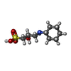
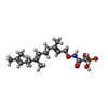


| #4: Chemical | ChemComp-EDO / Ethylene glycol Mass: 62.068 Da / Num. of mol.: 23 / Source method: obtained synthetically / Formula: C2H6O2 Mass: 62.068 Da / Num. of mol.: 23 / Source method: obtained synthetically / Formula: C2H6O2#5: Chemical | ChemComp-ZN / |  Mass: 65.409 Da / Num. of mol.: 1 / Source method: obtained synthetically / Formula: Zn Mass: 65.409 Da / Num. of mol.: 1 / Source method: obtained synthetically / Formula: Zn#6: Chemical |  Mass: 237.316 Da / Num. of mol.: 3 / Source method: obtained synthetically / Formula: C9H19NO4S Mass: 237.316 Da / Num. of mol.: 3 / Source method: obtained synthetically / Formula: C9H19NO4S#7: Chemical | ChemComp-FII / [( |  Mass: 359.398 Da / Num. of mol.: 1 / Source method: isolated from a natural source / Formula: C17H30NO5P / Feature type: SUBJECT OF INVESTIGATION Mass: 359.398 Da / Num. of mol.: 1 / Source method: isolated from a natural source / Formula: C17H30NO5P / Feature type: SUBJECT OF INVESTIGATION#8: Chemical | ChemComp-BT6 / | Thiophenol Mass: 110.177 Da / Num. of mol.: 1 / Source method: obtained synthetically / Formula: C6H6S Mass: 110.177 Da / Num. of mol.: 1 / Source method: obtained synthetically / Formula: C6H6S#9: Water | ChemComp-HOH / | WaterMass: 18.015 Da / Num. of mol.: 372 / Source method: isolated from a natural source / Formula: H2O |
|---|
-Details
| Has ligand of interest | Y |
|---|
-Experimental details
-Experiment
| Experiment | Method: X-RAY DIFFRACTION / Number of used crystals: 1 |
|---|
- Sample preparation
| Crystal | Density Matthews: 3.51 Å3/Da / Density % sol: 64.92 % |
|---|---|
| Crystal grow | Temperature: 290 K / Method: vapor diffusion, hanging drop / pH: 9.5 Details: 100mM CAPSO pH9.5, 50-75mM Li2SO4, 200mM NaCl, 16%-21% PEG4000. |
-Data collection
| Diffraction | Mean temperature: 100 K / Serial crystal experiment: N |
|---|---|
| Diffraction source | Source: SYNCHROTRON / Site: APS / Beamline: 22-ID / Wavelength: 1 Å |
| Detector | Type: DECTRIS EIGER X 16M / Detector: PIXEL / Date: Mar 31, 2023 |
| Radiation | Protocol: SINGLE WAVELENGTH / Monochromatic (M) / Laue (L): M / Scattering type: x-ray |
| Radiation wavelength | Wavelength: 1 Å / Relative weight: 1 |
| Reflection | Resolution: 1.892→49.77 Å / Num. obs: 103014 / % possible obs: 99.25 % / Redundancy: 5.7 % / CC1/2: 0.992 / Rmerge(I) obs: 0.117 / Rpim(I) all: 0.052 / Rrim(I) all: 0.129 / Net I/σ(I): 8.08 |
| Reflection shell | Resolution: 1.892→1.959 Å / Redundancy: 5.9 % / Rmerge(I) obs: 0.688 / Mean I/σ(I) obs: 1.82 / Num. unique obs: 10141 / CC1/2: 0.931 / Rpim(I) all: 0.306 / Rrim(I) all: 0.755 / % possible all: 98.87 |
- Processing
| Software |
| |||||||||||||||||||||||||||||||||||||||||||||||||||||||||||||||||||||||||||||||||||||||||||||||||||||||||||||||||||||||||||||||||||||||||||||||||||||||||||||||||||||||||||||||||||||||||||||||||||||||||||||||||||||||||
|---|---|---|---|---|---|---|---|---|---|---|---|---|---|---|---|---|---|---|---|---|---|---|---|---|---|---|---|---|---|---|---|---|---|---|---|---|---|---|---|---|---|---|---|---|---|---|---|---|---|---|---|---|---|---|---|---|---|---|---|---|---|---|---|---|---|---|---|---|---|---|---|---|---|---|---|---|---|---|---|---|---|---|---|---|---|---|---|---|---|---|---|---|---|---|---|---|---|---|---|---|---|---|---|---|---|---|---|---|---|---|---|---|---|---|---|---|---|---|---|---|---|---|---|---|---|---|---|---|---|---|---|---|---|---|---|---|---|---|---|---|---|---|---|---|---|---|---|---|---|---|---|---|---|---|---|---|---|---|---|---|---|---|---|---|---|---|---|---|---|---|---|---|---|---|---|---|---|---|---|---|---|---|---|---|---|---|---|---|---|---|---|---|---|---|---|---|---|---|---|---|---|---|---|---|---|---|---|---|---|---|---|---|---|---|---|---|---|---|
| Refinement | Method to determine structure: MOLECULAR REPLACEMENT / Resolution: 1.892→49.77 Å / SU ML: 0.21 / Cross valid method: FREE R-VALUE / σ(F): 1.35 / Phase error: 22.9 / Stereochemistry target values: ML
| |||||||||||||||||||||||||||||||||||||||||||||||||||||||||||||||||||||||||||||||||||||||||||||||||||||||||||||||||||||||||||||||||||||||||||||||||||||||||||||||||||||||||||||||||||||||||||||||||||||||||||||||||||||||||
| Solvent computation | Shrinkage radii: 0.9 Å / VDW probe radii: 1.1 Å / Solvent model: FLAT BULK SOLVENT MODEL | |||||||||||||||||||||||||||||||||||||||||||||||||||||||||||||||||||||||||||||||||||||||||||||||||||||||||||||||||||||||||||||||||||||||||||||||||||||||||||||||||||||||||||||||||||||||||||||||||||||||||||||||||||||||||
| Refinement step | Cycle: LAST / Resolution: 1.892→49.77 Å
| |||||||||||||||||||||||||||||||||||||||||||||||||||||||||||||||||||||||||||||||||||||||||||||||||||||||||||||||||||||||||||||||||||||||||||||||||||||||||||||||||||||||||||||||||||||||||||||||||||||||||||||||||||||||||
| Refine LS restraints |
| |||||||||||||||||||||||||||||||||||||||||||||||||||||||||||||||||||||||||||||||||||||||||||||||||||||||||||||||||||||||||||||||||||||||||||||||||||||||||||||||||||||||||||||||||||||||||||||||||||||||||||||||||||||||||
| LS refinement shell |
|
