 Movie
Movie Controller

[English] 日本語
 Yorodumi
- PDB-8t4t: Crystal structure of LC3A in complex with the LIR of TP53INP2/DOR -
+ Open data

- Basic information
| Entry | Database: PDB / ID: 8t4t | ||||||
|---|---|---|---|---|---|---|---|
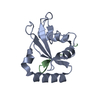
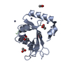
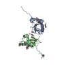
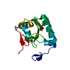
| Title | Crystal structure of LC3A in complex with the LIR of TP53INP2/DOR | ||||||
 Components Components | Tumor protein p53-inducible nuclear protein 2,Microtubule-associated proteins 1A/1B light chain 3A chimera Neoplasm Neoplasm | ||||||
 Keywords Keywords | PROTEIN BINDING / LC3A / Autophagy / DOR LIR / TP53INP2 LIR | ||||||
| Function / homology |  Function and homology information Function and homology informationnegative regulation of protein localization / cellular response to oxygen-glucose deprivation / autophagy of mitochondrion / tissue homeostasis / cellular response to nitrogen starvation / SMAD protein signal transduction / response to iron(II) ion / autolysosome / Macroautophagy / Receptor Mediated Mitophagy ...negative regulation of protein localization / cellular response to oxygen-glucose deprivation / autophagy of mitochondrion / tissue homeostasis / cellular response to nitrogen starvation / SMAD protein signal transduction / response to iron(II) ion / autolysosome / Macroautophagy / Receptor Mediated Mitophagy / p38MAPK cascade / autophagosome membrane / organelle membrane / autophagosome maturation / autophagosome assembly / autophagosome / JNK cascade / cellular response to copper ion / PINK1-PRKN Mediated Mitophagy / cellular response to amino acid starvation / cellular response to starvation / ubiquitin binding / macroautophagy / response to lead ion / protein localization / phospholipid binding / PML body / cellular response to hydrogen peroxide / osteoblast differentiation / late endosome / cytoplasmic vesicle / ubiquitin-dependent protein catabolic process / microtubule binding / microtubule / intracellular membrane-bounded organelle / glutamatergic synapse / ubiquitin protein ligase binding / positive regulation of DNA-templated transcription / nucleus / cytosolSimilarity search - Function | ||||||
| Biological species |  Homo sapiens (human) Homo sapiens (human) | ||||||
| Method | X-RAY DIFFRACTION / SYNCHROTRON / MOLECULAR REPLACEMENT / Resolution: 2.359 Å | ||||||
 Authors Authors | Ali, M.G.H. / Wahba, H.M. / Cyr, N. / Omichinski, J.G. | ||||||
| Funding support |  Canada, 1items Canada, 1items
| ||||||
 Citation Citation | Journal: Autophagy / Year: 2024 Title: Structural and functional characterization of the role of acetylation on the interactions of the human Atg8-family proteins with the autophagy receptor TP53INP2/DOR. Authors: Ali, M.G. / Wahba, H.M. / Igelmann, S. / Cyr, N. / Ferbeyre, G. / Omichinski, J.G. | ||||||
| History |
|
- Structure visualization
| Structure viewer | Molecule: MolmilJmol/JSmol |
|---|
- Downloads & links
-Download
| PDBx/mmCIF format | 8t4t.cif.gz | 119.1 KB | Display | PDBx/mmCIF format |
|---|---|---|---|---|
| PDB format | pdb8t4t.ent.gz | 93 KB | Display | PDB format |
| PDBx/mmJSON format | 8t4t.json.gz | Tree view | PDBx/mmJSON format | |
| Others |  Other downloads Other downloads |
-Validation report
| Arichive directory | https://data.pdbj.org/pub/pdb/validation_reports/t4/8t4tftp://data.pdbj.org/pub/pdb/validation_reports/t4/8t4t | HTTPS FTP |
|---|
-Related structure data

-Links
 PDBj
PDBj


- Assembly
| Deposited unit | 
| ||||||||
|---|---|---|---|---|---|---|---|---|---|
| 1 | 
| ||||||||
| 2 | 
| ||||||||
| Unit cell |
|
-Components
| #1: Protein | Neoplasm / Diabetes and obesity-regulated gene / p53-inducible protein U / PIG-U / Autophagy-related protein ...Diabetes and obesity-regulated gene / p53-inducible protein U / PIG-U / Autophagy-related protein LC3 A / Autophagy-related ubiquitin-like modifier LC3 A / MAP1 light chain 3-like protein 1 / MAP1A/MAP1B light chain 3 A / MAP1A/MAP1B LC3 A / Microtubule-associated protein 1 light chain 3 alpha Mass: 15722.941 Da / Num. of mol.: 2 Source method: isolated from a genetically manipulated source Source: (gene. exp.) Homo sapiens (human) / Gene: TP53INP2, C20orf110, DOR, PINH, MAP1LC3A / Production host:  Escherichia coli BL21(DE3) (bacteria) / References: UniProt: Q8IXH6, UniProt: Q9H492 Escherichia coli BL21(DE3) (bacteria) / References: UniProt: Q8IXH6, UniProt: Q9H492#2: Water | ChemComp-HOH / | Water Mass: 18.015 Da / Num. of mol.: 10 / Source method: isolated from a natural source / Formula: H2O Mass: 18.015 Da / Num. of mol.: 10 / Source method: isolated from a natural source / Formula: H2O |
|---|
-Experimental details
-Experiment
| Experiment | Method: X-RAY DIFFRACTION / Number of used crystals: 1 |
|---|
- Sample preparation
| Crystal | Density Matthews: 2.04 Å3/Da / Density % sol: 39.66 % |
|---|---|
| Crystal grow | Temperature: 293 K / Method: vapor diffusion, hanging drop Details: 12.5% PEG3350, 0.2M NH4-citrate, 5% isopropanol, 0.1M MES pH6.5 |
-Data collection
| Diffraction | Mean temperature: 100 K / Serial crystal experiment: N |
|---|---|
| Diffraction source | Source: SYNCHROTRON / Site: CLSI / Beamline: 08ID-1 / Wavelength: 0.97949 Å |
| Detector | Type: DECTRIS PILATUS3 6M / Detector: PIXEL / Date: Dec 16, 2017 |
| Radiation | Protocol: SINGLE WAVELENGTH / Monochromatic (M) / Laue (L): M / Scattering type: x-ray |
| Radiation wavelength | Wavelength: 0.97949 Å / Relative weight: 1 |
| Reflection | Resolution: 2.359→39.883 Å / Num. obs: 9618 / % possible obs: 89.74 % / Redundancy: 3 % / CC1/2: 0.995 / Net I/σ(I): 7.91 |
| Reflection shell | Resolution: 2.359→2.444 Å / Num. unique obs: 508 / CC1/2: 0.123 |
- Processing
| Software |
| ||||||||||||||||||||||||||||||||||||||||||||||||||||||||||||||||||||||||||||||||||||||||||||||||||||||||||||||||||||||||||||||||||||||||||||||||||||||||||||||||||||||||||||||||||||||||||||||||||||||||||||||||||||||||||||||||||||||||||||||||||||||||||||||||||||||||||||||||||||||||||||||||||||||||||||
|---|---|---|---|---|---|---|---|---|---|---|---|---|---|---|---|---|---|---|---|---|---|---|---|---|---|---|---|---|---|---|---|---|---|---|---|---|---|---|---|---|---|---|---|---|---|---|---|---|---|---|---|---|---|---|---|---|---|---|---|---|---|---|---|---|---|---|---|---|---|---|---|---|---|---|---|---|---|---|---|---|---|---|---|---|---|---|---|---|---|---|---|---|---|---|---|---|---|---|---|---|---|---|---|---|---|---|---|---|---|---|---|---|---|---|---|---|---|---|---|---|---|---|---|---|---|---|---|---|---|---|---|---|---|---|---|---|---|---|---|---|---|---|---|---|---|---|---|---|---|---|---|---|---|---|---|---|---|---|---|---|---|---|---|---|---|---|---|---|---|---|---|---|---|---|---|---|---|---|---|---|---|---|---|---|---|---|---|---|---|---|---|---|---|---|---|---|---|---|---|---|---|---|---|---|---|---|---|---|---|---|---|---|---|---|---|---|---|---|---|---|---|---|---|---|---|---|---|---|---|---|---|---|---|---|---|---|---|---|---|---|---|---|---|---|---|---|---|---|---|---|---|---|---|---|---|---|---|---|---|---|---|---|---|---|---|---|---|---|---|---|---|---|---|---|---|---|---|---|---|---|---|---|---|---|---|---|---|---|---|---|---|---|---|---|---|---|---|---|---|---|---|
| Refinement | Method to determine structure: MOLECULAR REPLACEMENT / Resolution: 2.359→39.883 Å / SU ML: 0.44 / Cross valid method: FREE R-VALUE / σ(F): 1.34 / Phase error: 35.89 / Stereochemistry target values: ML
| ||||||||||||||||||||||||||||||||||||||||||||||||||||||||||||||||||||||||||||||||||||||||||||||||||||||||||||||||||||||||||||||||||||||||||||||||||||||||||||||||||||||||||||||||||||||||||||||||||||||||||||||||||||||||||||||||||||||||||||||||||||||||||||||||||||||||||||||||||||||||||||||||||||||||||||
| Solvent computation | Shrinkage radii: 0.9 Å / VDW probe radii: 1.11 Å / Solvent model: FLAT BULK SOLVENT MODEL | ||||||||||||||||||||||||||||||||||||||||||||||||||||||||||||||||||||||||||||||||||||||||||||||||||||||||||||||||||||||||||||||||||||||||||||||||||||||||||||||||||||||||||||||||||||||||||||||||||||||||||||||||||||||||||||||||||||||||||||||||||||||||||||||||||||||||||||||||||||||||||||||||||||||||||||
| Refinement step | Cycle: LAST / Resolution: 2.359→39.883 Å
| ||||||||||||||||||||||||||||||||||||||||||||||||||||||||||||||||||||||||||||||||||||||||||||||||||||||||||||||||||||||||||||||||||||||||||||||||||||||||||||||||||||||||||||||||||||||||||||||||||||||||||||||||||||||||||||||||||||||||||||||||||||||||||||||||||||||||||||||||||||||||||||||||||||||||||||
| Refine LS restraints |
| ||||||||||||||||||||||||||||||||||||||||||||||||||||||||||||||||||||||||||||||||||||||||||||||||||||||||||||||||||||||||||||||||||||||||||||||||||||||||||||||||||||||||||||||||||||||||||||||||||||||||||||||||||||||||||||||||||||||||||||||||||||||||||||||||||||||||||||||||||||||||||||||||||||||||||||
| LS refinement shell |
| ||||||||||||||||||||||||||||||||||||||||||||||||||||||||||||||||||||||||||||||||||||||||||||||||||||||||||||||||||||||||||||||||||||||||||||||||||||||||||||||||||||||||||||||||||||||||||||||||||||||||||||||||||||||||||||||||||||||||||||||||||||||||||||||||||||||||||||||||||||||||||||||||||||||||||||
| Refinement TLS params. | Method: refined / Refine-ID: X-RAY DIFFRACTION
| ||||||||||||||||||||||||||||||||||||||||||||||||||||||||||||||||||||||||||||||||||||||||||||||||||||||||||||||||||||||||||||||||||||||||||||||||||||||||||||||||||||||||||||||||||||||||||||||||||||||||||||||||||||||||||||||||||||||||||||||||||||||||||||||||||||||||||||||||||||||||||||||||||||||||||||
| Refinement TLS group |
|
