 Movie
Movie Controller


+ Open data

- Basic information
| Entry | Database: PDB / ID: 8sig | ||||||
|---|---|---|---|---|---|---|---|
| Title | Crystal Structure of PRMT4 with Compound YD1-288 | ||||||
 Components Components | Histone-arginine methyltransferase CARM1 | ||||||
 Keywords Keywords | TRANSFERASE/INHIBITOR / PRMT4 / YD1-288 /  TRANSFERASE / Structural Genomics / PSI-2 / Protein Structure Initiative / Structural Genomics Consortium / SGC / TRANSFERASE-INHIBITOR complex TRANSFERASE / Structural Genomics / PSI-2 / Protein Structure Initiative / Structural Genomics Consortium / SGC / TRANSFERASE-INHIBITOR complex | ||||||
| Function / homology |  Function and homology information Function and homology information: / histone H3R17 methyltransferase activity / histone H3R2 methyltransferase activity / negative regulation of dendrite development / type I protein arginine methyltransferase / protein-arginine omega-N asymmetric methyltransferase activity / protein methyltransferase activity / histone arginine N-methyltransferase activity / regulation of intracellular estrogen receptor signaling pathway / replication fork reversal ...: / histone H3R17 methyltransferase activity / histone H3R2 methyltransferase activity / negative regulation of dendrite development / type I protein arginine methyltransferase / protein-arginine omega-N asymmetric methyltransferase activity / protein methyltransferase activity / histone arginine N-methyltransferase activity / regulation of intracellular estrogen receptor signaling pathway / replication fork reversal / protein-arginine N-methyltransferase activity / positive regulation of epithelial cell apoptotic process / histone methyltransferase activity / positive regulation of transcription by RNA polymerase I / nuclear replication fork / positive regulation of fat cell differentiation / response to cAMP / RORA activates gene expression / Regulation of lipid metabolism by PPARalpha / TP53 Regulates Transcription of Genes Involved in G2 Cell Cycle Arrest / BMAL1:CLOCK,NPAS2 activates circadian gene expression / Activation of gene expression by SREBF (SREBP) / nuclear receptor coactivator activity / lysine-acetylated histone binding / Heme signaling / Transcriptional activation of mitochondrial biogenesis / PPARA activates gene expression / Cytoprotection by HMOX1 / beta-catenin binding / RMTs methylate histone arginines / Transcriptional regulation of white adipocyte differentiation / Circadian Clock / DNA-binding transcription factor binding / Estrogen-dependent gene expression / transcription coactivator activity / transcription cis-regulatory region binding / positive regulation of cell population proliferation / regulation of DNA-templated transcription / nucleoplasm / nucleus / cytosol / cytoplasmSimilarity search - Function | ||||||
| Biological species |  Homo sapiens (human) Homo sapiens (human) | ||||||
| Method | X-RAY DIFFRACTION / SYNCHROTRON / MOLECULAR REPLACEMENT / Resolution: 1.78 Å | ||||||
 Authors Authors | Song, X. / Dong, A. / Deng, Y. / Huang, R. / Arrowsmith, C.H. / Edwards, A.M. / Min, J. / Structural Genomics Consortium (SGC) | ||||||
| Funding support |  Canada, 1items Canada, 1items
| ||||||
 Citation Citation | Journal: To be published Title: Crystal Structure of PRMT4 with Compound YD1-288 Authors: Song, X. / Dong, A. / Deng, Y. / Huang, R. / Arrowsmith, C.H. / Edwards, A.M. / Min, J. / Structural Genomics Consortium (SGC) | ||||||
| History |
|
- Structure visualization
| Structure viewer | Molecule: MolmilJmol/JSmol |
|---|
- Downloads & links
-Download
| PDBx/mmCIF format | 8sig.cif.gz | 94.8 KB | Display | PDBx/mmCIF format |
|---|---|---|---|---|
| PDB format | pdb8sig.ent.gz | 68 KB | Display | PDB format |
| PDBx/mmJSON format | 8sig.json.gz | Tree view | PDBx/mmJSON format | |
| Others |  Other downloads Other downloads |
-Validation report
| Arichive directory | https://data.pdbj.org/pub/pdb/validation_reports/si/8sigftp://data.pdbj.org/pub/pdb/validation_reports/si/8sig | HTTPS FTP |
|---|
-Related structure data
| Similar structure data |
|---|
-Links
 PDBj
PDBj


- Assembly
| Deposited unit | 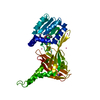
| ||||||||
|---|---|---|---|---|---|---|---|---|---|
| 1 | 
| ||||||||
| Unit cell |
| ||||||||
| Components on special symmetry positions |
| ||||||||
| Details | This construct only comprises the methyltransferase domain of CARM1 (PRMT4). The authors do not know the quaternary structure of this domain in isolation. However, the software PISA predicts it to be a dimer. |
-Components
| #1: Protein | Mass: 40946.520 Da / Num. of mol.: 1 / Fragment: methyltransferase domain Source method: isolated from a genetically manipulated source Source: (gene. exp.) Homo sapiens (human) / Gene: CARM1, PRMT4 / Production host:   Spodoptera frugiperda (fall armyworm) Spodoptera frugiperda (fall armyworm)References: UniProt: Q86X55, type I protein arginine methyltransferase | ||||
|---|---|---|---|---|---|
| #2: Chemical | ChemComp-GWU / Mass: 499.566 Da / Num. of mol.: 1 / Source method: obtained synthetically / Formula: C23H33N9O4 / Feature type: SUBJECT OF INVESTIGATION | ||||
| #3: Chemical | ChemComp-NA /   Mass: 22.990 Da / Num. of mol.: 1 / Source method: obtained synthetically / Formula: Na Mass: 22.990 Da / Num. of mol.: 1 / Source method: obtained synthetically / Formula: Na | ||||
| #4: Chemical | ChemComp-UNX /   Num. of mol.: 7 / Source method: obtained synthetically Num. of mol.: 7 / Source method: obtained synthetically#5: Water | ChemComp-HOH / | Water Mass: 18.015 Da / Num. of mol.: 248 / Source method: isolated from a natural source / Formula: H2O Mass: 18.015 Da / Num. of mol.: 248 / Source method: isolated from a natural source / Formula: H2OHas ligand of interest | Y | |
-Experimental details
-Experiment
| Experiment | Method: X-RAY DIFFRACTION / Number of used crystals: 1 |
|---|
- Sample preparation
| Crystal | Density Matthews: 3.32 Å3/Da / Density % sol: 62.98 % |
|---|---|
| Crystal grow | Temperature: 291 K / Method: vapor diffusion, sitting drop / pH: 7.5 Details: 0.2 M Calcium chloride dihydrate, 0.1 M HEPES sodium pH 7.5, 28% v/v Polyethylene glycol 400 |
-Data collection
| Diffraction | Mean temperature: 100 K / Serial crystal experiment: N | |||||||||||||||||||||||||||||||||||||||||||||||||||||||||||||||||||||||||||||||||||||||||||||||||||||||||||||||||||||||||||||||||||||||||||||||||||||||||||||||||||||||||||||||||||||||||||||
|---|---|---|---|---|---|---|---|---|---|---|---|---|---|---|---|---|---|---|---|---|---|---|---|---|---|---|---|---|---|---|---|---|---|---|---|---|---|---|---|---|---|---|---|---|---|---|---|---|---|---|---|---|---|---|---|---|---|---|---|---|---|---|---|---|---|---|---|---|---|---|---|---|---|---|---|---|---|---|---|---|---|---|---|---|---|---|---|---|---|---|---|---|---|---|---|---|---|---|---|---|---|---|---|---|---|---|---|---|---|---|---|---|---|---|---|---|---|---|---|---|---|---|---|---|---|---|---|---|---|---|---|---|---|---|---|---|---|---|---|---|---|---|---|---|---|---|---|---|---|---|---|---|---|---|---|---|---|---|---|---|---|---|---|---|---|---|---|---|---|---|---|---|---|---|---|---|---|---|---|---|---|---|---|---|---|---|---|---|---|---|
| Diffraction source | Source: SYNCHROTRON / Site: APS  / Beamline: 24-ID-E / Wavelength: 0.97918 Å / Beamline: 24-ID-E / Wavelength: 0.97918 Å | |||||||||||||||||||||||||||||||||||||||||||||||||||||||||||||||||||||||||||||||||||||||||||||||||||||||||||||||||||||||||||||||||||||||||||||||||||||||||||||||||||||||||||||||||||||||||||||
| Detector | Type: DECTRIS EIGER X 16M / Detector: PIXEL / Date: Sep 27, 2022 | |||||||||||||||||||||||||||||||||||||||||||||||||||||||||||||||||||||||||||||||||||||||||||||||||||||||||||||||||||||||||||||||||||||||||||||||||||||||||||||||||||||||||||||||||||||||||||||
| Radiation | Protocol: SINGLE WAVELENGTH / Monochromatic (M) / Laue (L): M / Scattering type: x-ray | |||||||||||||||||||||||||||||||||||||||||||||||||||||||||||||||||||||||||||||||||||||||||||||||||||||||||||||||||||||||||||||||||||||||||||||||||||||||||||||||||||||||||||||||||||||||||||||
| Radiation wavelength | Wavelength: 0.97918 Å / Relative weight: 1 | |||||||||||||||||||||||||||||||||||||||||||||||||||||||||||||||||||||||||||||||||||||||||||||||||||||||||||||||||||||||||||||||||||||||||||||||||||||||||||||||||||||||||||||||||||||||||||||
| Reflection | Resolution: 1.78→50 Å / Num. obs: 53004 / % possible obs: 100 % / Redundancy: 10.4 % / CC1/2: 0.998 / CC star: 0.999 / Rmerge(I) obs: 0.078 / Rpim(I) all: 0.025 / Rrim(I) all: 0.082 / Χ2: 1.18 / Net I/σ(I): 7.6 / Num. measured all: 553802 | |||||||||||||||||||||||||||||||||||||||||||||||||||||||||||||||||||||||||||||||||||||||||||||||||||||||||||||||||||||||||||||||||||||||||||||||||||||||||||||||||||||||||||||||||||||||||||||
| Reflection shell | Diffraction-ID: 1 / % possible all: 100
|
- Processing
| Software |
| ||||||||||||||||||||||||||||||||||||||||||||||||||||||||||||||||||||||||||||||||||||||||||||||||||||||||||||||||||||||||||||||||||||||||||||||||||||||||||||||||||||||||||||||||||||||
|---|---|---|---|---|---|---|---|---|---|---|---|---|---|---|---|---|---|---|---|---|---|---|---|---|---|---|---|---|---|---|---|---|---|---|---|---|---|---|---|---|---|---|---|---|---|---|---|---|---|---|---|---|---|---|---|---|---|---|---|---|---|---|---|---|---|---|---|---|---|---|---|---|---|---|---|---|---|---|---|---|---|---|---|---|---|---|---|---|---|---|---|---|---|---|---|---|---|---|---|---|---|---|---|---|---|---|---|---|---|---|---|---|---|---|---|---|---|---|---|---|---|---|---|---|---|---|---|---|---|---|---|---|---|---|---|---|---|---|---|---|---|---|---|---|---|---|---|---|---|---|---|---|---|---|---|---|---|---|---|---|---|---|---|---|---|---|---|---|---|---|---|---|---|---|---|---|---|---|---|---|---|---|---|
| Refinement | Method to determine structure: MOLECULAR REPLACEMENT / Resolution: 1.78→48.91 Å / Cor.coef. Fo:Fc: 0.964 / Cor.coef. Fo:Fc free: 0.961 / SU B: 2.259 / SU ML: 0.069 / Cross valid method: THROUGHOUT / ESU R: 0.091 / ESU R Free: 0.087 / Stereochemistry target values: MAXIMUM LIKELIHOOD / Details: HYDROGENS HAVE BEEN ADDED IN THE RIDING POSITIONS
| ||||||||||||||||||||||||||||||||||||||||||||||||||||||||||||||||||||||||||||||||||||||||||||||||||||||||||||||||||||||||||||||||||||||||||||||||||||||||||||||||||||||||||||||||||||||
| Solvent computation | Ion probe radii: 0.8 Å / Shrinkage radii: 0.8 Å / VDW probe radii: 1.2 Å / Solvent model: MASK | ||||||||||||||||||||||||||||||||||||||||||||||||||||||||||||||||||||||||||||||||||||||||||||||||||||||||||||||||||||||||||||||||||||||||||||||||||||||||||||||||||||||||||||||||||||||
| Displacement parameters | Biso mean: 27.756 Å2
| ||||||||||||||||||||||||||||||||||||||||||||||||||||||||||||||||||||||||||||||||||||||||||||||||||||||||||||||||||||||||||||||||||||||||||||||||||||||||||||||||||||||||||||||||||||||
| Refinement step | Cycle: 1 / Resolution: 1.78→48.91 Å
| ||||||||||||||||||||||||||||||||||||||||||||||||||||||||||||||||||||||||||||||||||||||||||||||||||||||||||||||||||||||||||||||||||||||||||||||||||||||||||||||||||||||||||||||||||||||
| Refine LS restraints |
|