 Movie
Movie Controller


+ Open data

- Basic information
| Entry | Database: PDB / ID: 8sfx | ||||||
|---|---|---|---|---|---|---|---|
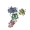
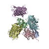
| Title | High Affinity nanobodies against GFP | ||||||
 Components Components |
| ||||||
 Keywords Keywords |  IMMUNE SYSTEM / Nanobody / nanobodies / GFP / green fluorescent protein / high-affinity antibody variant / antibody variant / single-domain antibody IMMUNE SYSTEM / Nanobody / nanobodies / GFP / green fluorescent protein / high-affinity antibody variant / antibody variant / single-domain antibody | ||||||
| Function / homology | Green fluorescent protein, GFP / Green fluorescent protein-related / Green fluorescent protein / Green fluorescent protein / bioluminescence / generation of precursor metabolites and energy / D-MALATE / PHOSPHATE ION / Green fluorescent protein Function and homology information Function and homology information | ||||||
| Biological species |   Aequorea victoria (jellyfish) Aequorea victoria (jellyfish) Lama glama (llama) Lama glama (llama) | ||||||
| Method | X-RAY DIFFRACTION / SYNCHROTRON / MOLECULAR REPLACEMENT / Resolution: 1.95 Å | ||||||
 Authors Authors | Ketaren, N.E. / Rout, M.P. / Bonanno, J.B. / Almo, S.C. | ||||||
| Funding support |  United States, 1items United States, 1items
| ||||||
 Citation Citation | Journal: To Be Published Title: High Affinity nanobodies against GFP Authors: Ketaren, N.E. / Bonanno, J.B. | ||||||
| History |
|
- Structure visualization
| Structure viewer | Molecule: MolmilJmol/JSmol |
|---|
- Downloads & links
-Download
| PDBx/mmCIF format | 8sfx.cif.gz | 411.1 KB | Display | PDBx/mmCIF format |
|---|---|---|---|---|
| PDB format | pdb8sfx.ent.gz | 340.6 KB | Display | PDB format |
| PDBx/mmJSON format | 8sfx.json.gz | Tree view | PDBx/mmJSON format | |
| Others |  Other downloads Other downloads |
-Validation report
| Arichive directory | https://data.pdbj.org/pub/pdb/validation_reports/sf/8sfxftp://data.pdbj.org/pub/pdb/validation_reports/sf/8sfx | HTTPS FTP |
|---|
-Related structure data


-Links
 PDBj
PDBj


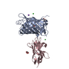
- Assembly
| Deposited unit | 
| |||||||||
|---|---|---|---|---|---|---|---|---|---|---|
| 1 |
| |||||||||
| 2 |
| |||||||||
| Unit cell |
| |||||||||
| Components on special symmetry positions |
|
-Components
-Protein , 2 types, 4 molecules ABDC
| #1: Protein | Mass: 28794.396 Da / Num. of mol.: 2 Source method: isolated from a genetically manipulated source Source: (gene. exp.) Aequorea victoria (jellyfish) / Gene: GFP / Production host:  Escherichia coli (E. coli) / References: UniProt: P42212 Escherichia coli (E. coli) / References: UniProt: P42212#2: Protein | Mass: 15470.181 Da / Num. of mol.: 2 Source method: isolated from a genetically manipulated source Source: (gene. exp.) Lama glama (llama) / Production host: Escherichia coli (E. coli) |
|---|
-Non-polymers , 5 types, 179 molecules 


| #3: Chemical | ChemComp-GOL / Glycerol Mass: 92.094 Da / Num. of mol.: 5 / Source method: obtained synthetically / Formula: C3H8O3 Mass: 92.094 Da / Num. of mol.: 5 / Source method: obtained synthetically / Formula: C3H8O3#4: Chemical | Malic acid Mass: 134.087 Da / Num. of mol.: 2 / Source method: obtained synthetically / Formula: C4H6O5 Mass: 134.087 Da / Num. of mol.: 2 / Source method: obtained synthetically / Formula: C4H6O5#5: Chemical | ChemComp-NA /  Mass: 22.990 Da / Num. of mol.: 19 / Source method: obtained synthetically / Formula: Na Mass: 22.990 Da / Num. of mol.: 19 / Source method: obtained synthetically / Formula: Na#6: Chemical | ChemComp-PO4 / Phosphate Mass: 94.971 Da / Num. of mol.: 7 / Source method: obtained synthetically / Formula: PO4 Mass: 94.971 Da / Num. of mol.: 7 / Source method: obtained synthetically / Formula: PO4#7: Water | ChemComp-HOH / | WaterMass: 18.015 Da / Num. of mol.: 146 / Source method: isolated from a natural source / Formula: H2O |
|---|
-Details
| Has ligand of interest | N |
|---|
-Experimental details
-Experiment
| Experiment | Method: X-RAY DIFFRACTION / Number of used crystals: 1 |
|---|
- Sample preparation
| Crystal | Density Matthews: 3.39 Å3/Da / Density % sol: 63.67 % |
|---|---|
| Crystal grow | Temperature: 291 K / Method: vapor diffusion, sitting drop Details: 1 M bis-tris propane pH 7.0, 1.2 M DL-malic acid pH 7.0 |
-Data collection
| Diffraction | Mean temperature: 100 K / Serial crystal experiment: N |
|---|---|
| Diffraction source | Source: SYNCHROTRON / Site: APS / Beamline: 31-ID / Wavelength: 0.9793 Å |
| Detector | Type: RAYONIX MX225HE / Detector: CCD / Date: Nov 12, 2012 |
| Radiation | Protocol: SINGLE WAVELENGTH / Monochromatic (M) / Laue (L): M / Scattering type: x-ray |
| Radiation wavelength | Wavelength: 0.9793 Å / Relative weight: 1 |
| Reflection | Resolution: 1.95→100 Å / Num. obs: 85311 / % possible obs: 100 % / Redundancy: 15.1 % / Rmerge(I) obs: 0.13 / Net I/σ(I): 11.1 |
| Reflection shell | Resolution: 1.95→2.06 Å / Rmerge(I) obs: 1.403 / Mean I/σ(I) obs: 2 / Num. unique obs: 12451 / % possible all: 100 |
- Processing
| Software |
| |||||||||||||||||||||||||||||||||||||||||||||||||||||||||||||||||||||||||||||||||||||||||||||||||||||||||
|---|---|---|---|---|---|---|---|---|---|---|---|---|---|---|---|---|---|---|---|---|---|---|---|---|---|---|---|---|---|---|---|---|---|---|---|---|---|---|---|---|---|---|---|---|---|---|---|---|---|---|---|---|---|---|---|---|---|---|---|---|---|---|---|---|---|---|---|---|---|---|---|---|---|---|---|---|---|---|---|---|---|---|---|---|---|---|---|---|---|---|---|---|---|---|---|---|---|---|---|---|---|---|---|---|---|---|
| Refinement | Method to determine structure: MOLECULAR REPLACEMENT / Resolution: 1.95→36.67 Å / SU ML: 0.23 / Cross valid method: FREE R-VALUE / σ(F): 0.01 / Phase error: 27.62 / Stereochemistry target values: ML
| |||||||||||||||||||||||||||||||||||||||||||||||||||||||||||||||||||||||||||||||||||||||||||||||||||||||||
| Solvent computation | Shrinkage radii: 0.9 Å / VDW probe radii: 1.11 Å / Solvent model: FLAT BULK SOLVENT MODEL | |||||||||||||||||||||||||||||||||||||||||||||||||||||||||||||||||||||||||||||||||||||||||||||||||||||||||
| Refinement step | Cycle: LAST / Resolution: 1.95→36.67 Å
| |||||||||||||||||||||||||||||||||||||||||||||||||||||||||||||||||||||||||||||||||||||||||||||||||||||||||
| Refine LS restraints |
| |||||||||||||||||||||||||||||||||||||||||||||||||||||||||||||||||||||||||||||||||||||||||||||||||||||||||
| LS refinement shell |
| |||||||||||||||||||||||||||||||||||||||||||||||||||||||||||||||||||||||||||||||||||||||||||||||||||||||||
| Refinement TLS params. | Method: refined / Origin x: -27.8072 Å / Origin y: -10.3191 Å / Origin z: 5.773 Å
| |||||||||||||||||||||||||||||||||||||||||||||||||||||||||||||||||||||||||||||||||||||||||||||||||||||||||
| Refinement TLS group | Selection details: all |
