 Movie
Movie Controller

[English] 日本語
 Yorodumi
- PDB-8ruc: ACTIVATED SPINACH RUBISCO COMPLEXED WITH 2-CARBOXYARABINITOL BISP... -
+ Open data

- Basic information
| Entry | Database: PDB / ID: 8ruc | |||||||||
|---|---|---|---|---|---|---|---|---|---|---|
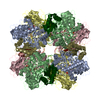
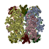
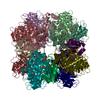
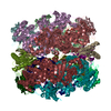
| Title | ACTIVATED SPINACH RUBISCO COMPLEXED WITH 2-CARBOXYARABINITOL BISPHOSPHATE | |||||||||
 Components Components | (RIBULOSE-1,5-BISPHOSPHATE CARBOXYLASE/OXYGENASE) x 2 | |||||||||
 Keywords Keywords | LYASE (CARBON-CARBON) /  PHOTOSYNTHESIS / CARBON DIOXIDE FIXATION / PHOTORESPIRATION / LYASE / OXIDOREDUCTASE / MONOOXYGENASE / CHLOROPLAST PHOTOSYNTHESIS / CARBON DIOXIDE FIXATION / PHOTORESPIRATION / LYASE / OXIDOREDUCTASE / MONOOXYGENASE / CHLOROPLAST | |||||||||
| Function / homology |  Function and homology informationphotorespiration / ribulose-bisphosphate carboxylase / ribulose-bisphosphate carboxylase activity / reductive pentose-phosphate cycle / chloroplast / monooxygenase activity / magnesium ion binding Function and homology informationphotorespiration / ribulose-bisphosphate carboxylase / ribulose-bisphosphate carboxylase activity / reductive pentose-phosphate cycle / chloroplast / monooxygenase activity / magnesium ion bindingSimilarity search - Function | |||||||||
| Biological species |  Spinacia oleracea (spinach) Spinacia oleracea (spinach) | |||||||||
| Method | X-RAY DIFFRACTION / SYNCHROTRON / MIR STARTING MODEL FOR MOLECULAR REPLACEMENT: NULL / Resolution: 1.6 Å | |||||||||
 Authors Authors | Andersson, I. / Knight, S. / Branden, C.-I. | |||||||||
 Citation Citation | Journal: J.Mol.Biol. / Year: 1996 Title: Large structures at high resolution: the 1.6 A crystal structure of spinach ribulose-1,5-bisphosphate carboxylase/oxygenase complexed with 2-carboxyarabinitol bisphosphate. Authors: Andersson, I. #1: Journal: J.Mol.Biol. / Year: 1990Title: Crystallographic Analysis of Ribulose 1,5-Bisphosphate Carboxylase from Spinach at 2.4 A Resolution Authors: Knight, S. / Andersson, I. / Branden, C.I. #2: Journal: Nature / Year: 1989Title: Crystal Structure of the Active Site of Ribulose-Bisphosphate Carboxylase Authors: Andersson, I. / Knight, S. / Schneider, G. / Lindqvist, Y. / Lundqvist, T. / Branden, C.-I. / Lorimer, G.H. #3: Journal: Science / Year: 1989Title: Reexamination of the Three-Dimensional Structure of the Small Subunit of Rubisco from Higher Plants Authors: Knight, S. / Andersson, I. / Branden, C.-I. | |||||||||
| History |
|
- Structure visualization
| Structure viewer | Molecule: MolmilJmol/JSmol |
|---|
- Downloads & links
-Download
| PDBx/mmCIF format | 8ruc.cif.gz | 493.2 KB | Display | PDBx/mmCIF format |
|---|---|---|---|---|
| PDB format | pdb8ruc.ent.gz | 401.8 KB | Display | PDB format |
| PDBx/mmJSON format | 8ruc.json.gz | Tree view | PDBx/mmJSON format | |
| Others |  Other downloads Other downloads |
-Validation report
| Arichive directory | https://data.pdbj.org/pub/pdb/validation_reports/ru/8rucftp://data.pdbj.org/pub/pdb/validation_reports/ru/8ruc | HTTPS FTP |
|---|
-Related structure data
| Similar structure data |
|---|


-Links
 PDBj
PDBj
- Assembly
| Deposited unit | 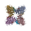
| ||||||||
|---|---|---|---|---|---|---|---|---|---|
| 1 | 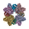
| ||||||||
| Unit cell |
| ||||||||
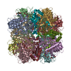
| Details | THE ENZYME IS A HEXADECAMER L8S8 OF EIGHT L (LARGE) SUBUNIT AND 8 S (SMALL) SUBUNITS. THE L SUBUNIT HAS BEEN ASSIGNED CHAIN IDENTIFIERS A, B, C, D, E, F, G, AND H. THE S SUBUNIT HAS BEEN ASSIGNED CHAIN IDENTIFIERS I, J, K, L, M, N, O, AND P. IN THE CATALYTICALLY COMPETENT MOLECULE, DIMERS OF THE LARGE SUBUNITS FORM FUNCTIONAL UNITS. IN REFERENCE 1 THESE DIMERS ARE DESIGNATED *AB*, *CD*, *EF*, AND *GH*. THIS ENTRY PRESENTS THE CRYSTALLOGRAPHIC ASYMMETRIC UNIT WHICH CONTAINS HALF THE MOLECULE: FOUR L AND FOUR S SUBUNITS RELATED BY A FOUR-FOLD AXIS THROUGH THE MOLECULAR CENTER AT X=0, Y=1/4, Z=1/4. THE FOUR L CHAINS PRESENT ARE A, C, E, G AND THE FOUR S CHAINS PRESENT ARE I, J, K, L. ALSO INCLUDED ARE FOUR MAGNESIUM IONS, FOUR INHIBITOR MOLECULES (CAP) AND 1520 WATERS. |
-Components
| #1: Protein | Mass: 52849.742 Da / Num. of mol.: 4 / Source method: isolated from a natural source / Source: (natural) Spinacia oleracea (spinach) / Organ: LEAFReferences: UniProt: P00875, ribulose-bisphosphate carboxylase#2: Protein | Mass: 14638.671 Da / Num. of mol.: 4 / Source method: isolated from a natural source / Source: (natural) Spinacia oleracea (spinach) / Organ: LEAFReferences: UniProt: P00870, ribulose-bisphosphate carboxylase#3: Chemical | ChemComp-MG /   Mass: 24.305 Da / Num. of mol.: 4 / Source method: obtained synthetically / Formula: Mg Mass: 24.305 Da / Num. of mol.: 4 / Source method: obtained synthetically / Formula: Mg#4: Sugar | ChemComp-CAP /   Type: saccharideCarbohydrate / Mass: 356.115 Da / Num. of mol.: 4 / Source method: obtained synthetically / Formula: C6H14O13P2 Type: saccharideCarbohydrate / Mass: 356.115 Da / Num. of mol.: 4 / Source method: obtained synthetically / Formula: C6H14O13P2#5: Water | ChemComp-HOH / | Water Mass: 18.015 Da / Num. of mol.: 1520 / Source method: isolated from a natural source / Formula: H2O Mass: 18.015 Da / Num. of mol.: 1520 / Source method: isolated from a natural source / Formula: H2OCompound details | RESIDUE 201 OF THE L SUBUNITS IS A MODIFIED ACTIVATOR LYSINE WHICH IS CARBAMYLATED AT THE EPSILON- ...RESIDUE 201 OF THE L SUBUNITS IS A MODIFIED ACTIVATOR LYSINE WHICH IS CARBAMYLAT | |
|---|
-Experimental details
-Experiment
| Experiment | Method: X-RAY DIFFRACTION / Number of used crystals: 2 |
|---|
- Sample preparation
| Crystal | Density Matthews: 2.3 Å3/Da / Density % sol: 40 % | ||||||||||||||||||||||||||||||||||||||||||
|---|---|---|---|---|---|---|---|---|---|---|---|---|---|---|---|---|---|---|---|---|---|---|---|---|---|---|---|---|---|---|---|---|---|---|---|---|---|---|---|---|---|---|---|
| Crystal grow | pH: 7.5 / Details: pH 7.5 | ||||||||||||||||||||||||||||||||||||||||||
| Crystal | *PLUS | ||||||||||||||||||||||||||||||||||||||||||
| Crystal grow | *PLUS Method: vapor diffusion, hanging drop / Details: Andersson, I., (1983) J. Biol. Chem., 258, 14088. / PH range low: 6 / PH range high: 5 | ||||||||||||||||||||||||||||||||||||||||||
| Components of the solutions | *PLUS
|
-Data collection
| Diffraction | Mean temperature: 283 K |
|---|---|
| Diffraction source | Source: SYNCHROTRON / Site: Photon Factory  / Beamline: BL-6A / Wavelength: 1.04 / Beamline: BL-6A / Wavelength: 1.04 |
| Detector | Type: RIGAKU RAXIS IV++ / Detector: IMAGE PLATE / Date: Jul 7, 1990 |
| Radiation | Monochromatic (M) / Laue (L): M / Scattering type: x-ray |
| Radiation wavelength | Wavelength: 1.04 Å / Relative weight: 1 |
| Reflection | Resolution: 1.5→20 Å / Num. obs: 277449 / Redundancy: 4 % / Rmerge(I) obs: 0.069 |
| Reflection shell | Resolution: 1.6→1.67 Å / % possible all: 49 |
| Reflection | *PLUS Num. measured all: 1131480 |
- Processing
| Software |
| ||||||||||||||||||||||||||||||||||||||||||||||||||||||||||||
|---|---|---|---|---|---|---|---|---|---|---|---|---|---|---|---|---|---|---|---|---|---|---|---|---|---|---|---|---|---|---|---|---|---|---|---|---|---|---|---|---|---|---|---|---|---|---|---|---|---|---|---|---|---|---|---|---|---|---|---|---|---|
| Refinement | Method to determine structure: MIR STARTING MODEL FOR MOLECULAR REPLACEMENT: NULL Resolution: 1.6→7 Å / σ(F): 0 Details: NO ELECTRON DENSITY IS OBSERVED FOR THE FIRST EIGHT RESIDUES OF THE LARGE SUBUNIT. NO COORDINATES ARE PRESENTED FOR THESE RESIDUES. THE ELECTRON DENSITY FOR RESIDUES ALA 9, SER 10 AND VAL 11 ...Details: NO ELECTRON DENSITY IS OBSERVED FOR THE FIRST EIGHT RESIDUES OF THE LARGE SUBUNIT. NO COORDINATES ARE PRESENTED FOR THESE RESIDUES. THE ELECTRON DENSITY FOR RESIDUES ALA 9, SER 10 AND VAL 11 OF THE L SUBUNITS IS WEAK.
| ||||||||||||||||||||||||||||||||||||||||||||||||||||||||||||
| Displacement parameters | Biso mean: 15.2 Å2 | ||||||||||||||||||||||||||||||||||||||||||||||||||||||||||||
| Refinement step | Cycle: LAST / Resolution: 1.6→7 Å
| ||||||||||||||||||||||||||||||||||||||||||||||||||||||||||||
| Refine LS restraints |
| ||||||||||||||||||||||||||||||||||||||||||||||||||||||||||||
| Software | *PLUS Name: X-PLOR / Version: 3.1 / Classification: refinement | ||||||||||||||||||||||||||||||||||||||||||||||||||||||||||||
| Refine LS restraints | *PLUS
|
