 Movie
Movie Controller

[English] 日本語
 Yorodumi
- PDB-8re2: Crystal Structure determination of Dye-decolorizing Peroxidase (D... -
+ Open data

- Basic information
| Entry | Database: PDB / ID: 8re2 | |||||||||||||||||||||
|---|---|---|---|---|---|---|---|---|---|---|---|---|---|---|---|---|---|---|---|---|---|---|
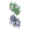
| Title | Crystal Structure determination of Dye-decolorizing Peroxidase (DyP) from Deinoccoccus radiodurans | |||||||||||||||||||||
 Components Components | Peroxidase, putative | |||||||||||||||||||||
 Keywords Keywords | STRUCTURAL PROTEIN / Heme protein / peroxidase / ferredoxin like fold / Hydrogen Peroxide | |||||||||||||||||||||
| Function / homology | DyP dimeric alpha+beta barrel domain / DyP-type peroxidase family. / Dyp-type peroxidase / Dimeric alpha-beta barrel / peroxidase activity / heme binding / cytosol / PROTOPORPHYRIN IX CONTAINING FE / Peroxidase, putative Function and homology information Function and homology information | |||||||||||||||||||||
| Biological species |  Deinococcus radiodurans (radioresistant) Deinococcus radiodurans (radioresistant) | |||||||||||||||||||||
| Method | X-RAY DIFFRACTION / SYNCHROTRON / MOLECULAR REPLACEMENT / Resolution: 2.2 Å | |||||||||||||||||||||
 Authors Authors | Salgueiro, B.A. / Frade, K. / Frazao, C. / Moe, E. | |||||||||||||||||||||
| Funding support |  Portugal, 6items Portugal, 6items
| |||||||||||||||||||||
 Citation Citation | Journal: Molecules / Year: 2024 Title: Biochemical, Biophysical, and Structural Analysis of an Unusual DyP from the Extremophile Deinococcus radiodurans. Authors: Frade, K. / Silveira, C.M. / Salgueiro, B.A. / Mendes, S. / Martins, L.O. / Frazao, C. / Todorovic, S. / Moe, E. | |||||||||||||||||||||
| History |
|
- Structure visualization
| Structure viewer | Molecule: MolmilJmol/JSmol |
|---|
- Downloads & links
-Download
| PDBx/mmCIF format | 8re2.cif.gz | 104.2 KB | Display | PDBx/mmCIF format |
|---|---|---|---|---|
| PDB format | pdb8re2.ent.gz | 76.8 KB | Display | PDB format |
| PDBx/mmJSON format | 8re2.json.gz | Tree view | PDBx/mmJSON format | |
| Others |  Other downloads Other downloads |
-Validation report
| Arichive directory | https://data.pdbj.org/pub/pdb/validation_reports/re/8re2ftp://data.pdbj.org/pub/pdb/validation_reports/re/8re2 | HTTPS FTP |
|---|
-Related structure data
-Links
 PDBj
PDBj- Assembly
| Deposited unit | 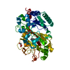
| ||||||||
|---|---|---|---|---|---|---|---|---|---|
| 1 |
| ||||||||
| Unit cell |
|
-Components
| #1: Protein | Mass: 49501.855 Da / Num. of mol.: 1 Source method: isolated from a genetically manipulated source Source: (gene. exp.) Deinococcus radiodurans (radioresistant)Gene: DR_A0145 / Production host: Escherichia coli BL21(DE3) (bacteria) / Variant (production host): * / References: UniProt: Q9RZ08 | ||||||
|---|---|---|---|---|---|---|---|
| #2: Chemical | ChemComp-HEM / Heme B  Mass: 616.487 Da / Num. of mol.: 1 / Source method: obtained synthetically / Formula: C34H32FeN4O4 / Feature type: SUBJECT OF INVESTIGATION Mass: 616.487 Da / Num. of mol.: 1 / Source method: obtained synthetically / Formula: C34H32FeN4O4 / Feature type: SUBJECT OF INVESTIGATION | ||||||
| #3: Chemical | Glycerol  Mass: 92.094 Da / Num. of mol.: 2 / Source method: obtained synthetically / Formula: C3H8O3 Mass: 92.094 Da / Num. of mol.: 2 / Source method: obtained synthetically / Formula: C3H8O3#4: Chemical |   Mass: 24.305 Da / Num. of mol.: 3 / Source method: obtained synthetically / Formula: Mg Mass: 24.305 Da / Num. of mol.: 3 / Source method: obtained synthetically / Formula: Mg#5: Water | ChemComp-HOH / | Water Mass: 18.015 Da / Num. of mol.: 49 / Source method: isolated from a natural source / Formula: H2O Mass: 18.015 Da / Num. of mol.: 49 / Source method: isolated from a natural source / Formula: H2OHas ligand of interest | Y | |
-Experimental details
-Experiment
| Experiment | Method: X-RAY DIFFRACTION / Number of used crystals: 1 |
|---|
- Sample preparation
| Crystal | Density Matthews: 2.67 Å3/Da / Density % sol: 53.93 % |
|---|---|
| Crystal grow | Temperature: 293.15 K / Method: vapor diffusion / pH: 8.5 / Details: 18% PEG 8000, 0.1 M CHES pH 8.5 |
-Data collection
| Diffraction | Mean temperature: 100 K / Serial crystal experiment: N |
|---|---|
| Diffraction source | Source: SYNCHROTRON / Site: Diamond  / Beamline: I03 / Wavelength: 0.97625 Å / Beamline: I03 / Wavelength: 0.97625 Å |
| Detector | Type: DECTRIS EIGER2 XE 16M / Detector: PIXEL / Date: Jul 18, 2017 |
| Radiation | Protocol: SINGLE WAVELENGTH / Monochromatic (M) / Laue (L): M / Scattering type: x-ray |
| Radiation wavelength | Wavelength: 0.97625 Å / Relative weight: 1 |
| Reflection | Resolution: 2.2→55.54 Å / Num. obs: 25843 / % possible obs: 99.5 % / Redundancy: 4.02 % / Biso Wilson estimate: 62.2 Å2 / Rpim(I) all: 0.0352 / Rsym value: 0.066 / Net I/σ(I): 10.96 |
| Reflection shell | Resolution: 2.2→2.33 Å / Num. unique obs: 4160 / Rpim(I) all: 0.347 / Rsym value: 1.08 |
- Processing
| Software |
| |||||||||||||||||||||||||||||||||||||||||||||||||||||||||||||||||||||||||||||||||||||||||||||||||||||||||
|---|---|---|---|---|---|---|---|---|---|---|---|---|---|---|---|---|---|---|---|---|---|---|---|---|---|---|---|---|---|---|---|---|---|---|---|---|---|---|---|---|---|---|---|---|---|---|---|---|---|---|---|---|---|---|---|---|---|---|---|---|---|---|---|---|---|---|---|---|---|---|---|---|---|---|---|---|---|---|---|---|---|---|---|---|---|---|---|---|---|---|---|---|---|---|---|---|---|---|---|---|---|---|---|---|---|---|
| Refinement | Method to determine structure: MOLECULAR REPLACEMENT / Resolution: 2.2→39.31 Å / SU ML: 0.34 / Cross valid method: FREE R-VALUE / σ(F): 1.93 / Phase error: 32.27 / Stereochemistry target values: ML
| |||||||||||||||||||||||||||||||||||||||||||||||||||||||||||||||||||||||||||||||||||||||||||||||||||||||||
| Solvent computation | Shrinkage radii: 0.9 Å / VDW probe radii: 1.11 Å / Solvent model: FLAT BULK SOLVENT MODEL | |||||||||||||||||||||||||||||||||||||||||||||||||||||||||||||||||||||||||||||||||||||||||||||||||||||||||
| Refinement step | Cycle: LAST / Resolution: 2.2→39.31 Å
| |||||||||||||||||||||||||||||||||||||||||||||||||||||||||||||||||||||||||||||||||||||||||||||||||||||||||
| Refine LS restraints |
| |||||||||||||||||||||||||||||||||||||||||||||||||||||||||||||||||||||||||||||||||||||||||||||||||||||||||
| LS refinement shell |
|
