National Institutes of Health/National Institute of Neurological Disorders and Stroke (NIH/NINDS)
NIH R01 NS115716
United States
Medical Research Council (MRC, United Kingdom)
MC_UP_1201/16
United Kingdom
Novo Nordisk Foundation
NNF17OC0030788
Denmark
Citation
Journal: Cell / Year: 2024 Title: PNMA2 forms immunogenic non-enveloped virus-like capsids associated with paraneoplastic neurological syndrome. Authors: Junjie Xu / Simon Erlendsson / Manvendra Singh / G Aaron Holling / Matthew Regier / Iosune Ibiricu / Jenifer Einstein / Michael P Hantak / Gregory S Day / Amanda L Piquet / Tammy L Smith / ...Authors: Junjie Xu / Simon Erlendsson / Manvendra Singh / G Aaron Holling / Matthew Regier / Iosune Ibiricu / Jenifer Einstein / Michael P Hantak / Gregory S Day / Amanda L Piquet / Tammy L Smith / Stacey L Clardy / Alexandra M Whiteley / Cédric Feschotte / John A G Briggs / Jason D Shepherd / Abstract: The paraneoplastic Ma antigen (PNMA) proteins are associated with cancer-induced paraneoplastic syndromes that present with an autoimmune response and neurological symptoms. Why PNMA proteins are ...The paraneoplastic Ma antigen (PNMA) proteins are associated with cancer-induced paraneoplastic syndromes that present with an autoimmune response and neurological symptoms. Why PNMA proteins are associated with this severe autoimmune disease is unclear. PNMA genes are predominantly expressed in the central nervous system and are ectopically expressed in some tumors. We show that PNMA2, which has been co-opted from a Ty3 retrotransposon, encodes a protein that is released from cells as non-enveloped virus-like capsids. Recombinant PNMA2 capsids injected into mice induce autoantibodies that preferentially bind external "spike" PNMA2 capsid epitopes, whereas a capsid-assembly-defective PNMA2 protein is not immunogenic. PNMA2 autoantibodies in cerebrospinal fluid of patients with anti-Ma2 paraneoplastic disease show similar preferential binding to spike capsid epitopes. PNMA2 capsid-injected mice develop learning and memory deficits. These observations suggest that PNMA2 capsids act as an extracellular antigen, capable of generating an autoimmune response that results in neurological deficits.
Average exposure time: 40 sec. / Electron dose: 40 e/Å2 / Detector mode: COUNTING / Film or detector model: FEI FALCON III (4k x 4k) / Num. of real images: 3005
Image scans
Width: 4096 / Height: 4096
-
Processing
EM software
ID
Name
Version
Category
1
RELION
4
particleselection
2
EPU
imageacquisition
4
CTFFIND
4.1
CTFcorrection
7
UCSF ChimeraX
1.15
modelfitting
10
RELION
4
finalEulerassignment
11
RELION
4
classification
12
RELION
4
3Dreconstruction
13
Coot
0.9
modelrefinement
14
PHENIX
1.20.1
modelrefinement
15
UCSF ChimeraX
1.15
modelrefinement
16
ISOLDE
modelrefinement
CTF correction
Type: PHASE FLIPPING AND AMPLITUDE CORRECTION
Particle selection
Num. of particles selected: 2125200
3D reconstruction
Resolution: 3.2 Å / Resolution method: FSC 0.143 CUT-OFF / Num. of particles: 835009 / Algorithm: FOURIER SPACE / Num. of class averages: 1 / Symmetry type: POINT
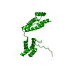
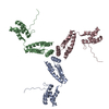
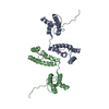
Atomic model building
B value: 105 / Protocol: OTHER / Space: REAL
Atomic model building
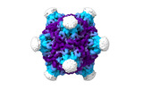
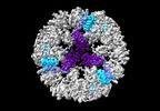
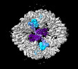
Accession code: 8RB3 / Details: Full capsid fitted model / Source name: Other / Type: experimental model
+
About Yorodumi
-
News
-
Feb 9, 2022. New format data for meta-information of EMDB entries
New format data for meta-information of EMDB entries
Version 3 of the EMDB header file is now the official format.
The previous official version 1.9 will be removed from the archive.
In the structure databanks used in Yorodumi, some data are registered as the other names, "COVID-19 virus" and "2019-nCoV". Here are the details of the virus and the list of structure data.
Jan 31, 2019. EMDB accession codes are about to change! (news from PDBe EMDB page)
EMDB accession codes are about to change! (news from PDBe EMDB page)
The allocation of 4 digits for EMDB accession codes will soon come to an end. Whilst these codes will remain in use, new EMDB accession codes will include an additional digit and will expand incrementally as the available range of codes is exhausted. The current 4-digit format prefixed with “EMD-” (i.e. EMD-XXXX) will advance to a 5-digit format (i.e. EMD-XXXXX), and so on. It is currently estimated that the 4-digit codes will be depleted around Spring 2019, at which point the 5-digit format will come into force.
The EM Navigator/Yorodumi systems omit the EMD- prefix.
Related info.:Q: What is EMD? / ID/Accession-code notation in Yorodumi/EM Navigator
Yorodumi is a browser for structure data from EMDB, PDB, SASBDB, etc.
This page is also the successor to EM Navigator detail page, and also detail information page/front-end page for Omokage search.
The word "yorodu" (or yorozu) is an old Japanese word meaning "ten thousand". "mi" (miru) is to see.
Related info.:EMDB / PDB / SASBDB / Comparison of 3 databanks / Yorodumi Search / Aug 31, 2016. New EM Navigator & Yorodumi / Yorodumi Papers / Jmol/JSmol / Function and homology information / Changes in new EM Navigator and Yorodumi
 Movie
Movie Controller


 Open data
 Basic information
 Components
 Keywords
 VIRUS LIKE PARTICLE / Endogenous retrovirus. PNMA2 / PNMA / Paraneoplastic syndrome / Paraneoplastic antigen Ma2 / VLP.
 Function and homology information
 Authors
 United States,
  United Kingdom,
  Denmark, 3items
  Citation
 Structure visualization
 Downloads & links
 Other downloads
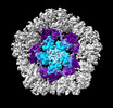
 PDBj
PDBj Assembly
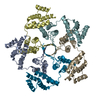

 Sample preparation
 Electron microscopy imaging
 Processing