 Movie
Movie Controller


+ Open data

- Basic information
| Entry | Database: PDB / ID: 8r37 | ||||||
|---|---|---|---|---|---|---|---|
| Title | Klebsiella pneumoniae fosfomycin-resistance protein (FosAKP) | ||||||
 Components Components | FosA family fosfomycin resistance glutathione transferase | ||||||
 Keywords Keywords |  TRANSFERASE / Antibiotic resistance / Klebsiella pneumoniae / Glutathione / Fosfomycin / metalloenzyme / biosensor TRANSFERASE / Antibiotic resistance / Klebsiella pneumoniae / Glutathione / Fosfomycin / metalloenzyme / biosensor | ||||||
| Function / homology |  Function and homology information Function and homology information | ||||||
| Biological species |  Klebsiella pneumoniae (bacteria) Klebsiella pneumoniae (bacteria) | ||||||
| Method | X-RAY DIFFRACTION / SYNCHROTRON / MOLECULAR REPLACEMENT / Resolution: 1.48 Å | ||||||
 Authors Authors | Papageorgiou, A.C. / Varotsou, C. / Labrou, N.E. | ||||||
| Funding support | 1items
| ||||||
 Citation Citation | Journal: Int J Mol Sci / Year: 2023 Title: Structural Studies of Klebsiella pneumoniae Fosfomycin-Resistance Protein and Its Application for the Development of an Optical Biosensor for Fosfomycin Determination. Authors: Varotsou, C. / Ataya, F. / Papageorgiou, A.C. / Labrou, N.E. | ||||||
| History |
|
- Structure visualization
| Structure viewer | Molecule: MolmilJmol/JSmol |
|---|
- Downloads & links
-Download
| PDBx/mmCIF format | 8r37.cif.gz | 97.4 KB | Display | PDBx/mmCIF format |
|---|---|---|---|---|
| PDB format | pdb8r37.ent.gz | 58.7 KB | Display | PDB format |
| PDBx/mmJSON format | 8r37.json.gz | Tree view | PDBx/mmJSON format | |
| Others |  Other downloads Other downloads |
-Validation report
| Arichive directory | https://data.pdbj.org/pub/pdb/validation_reports/r3/8r37ftp://data.pdbj.org/pub/pdb/validation_reports/r3/8r37 | HTTPS FTP |
|---|
-Related structure data
| Similar structure data |
|---|
-Links
 PDBj
PDBj


- Assembly
| Deposited unit | 
| ||||||||||||
|---|---|---|---|---|---|---|---|---|---|---|---|---|---|
| 1 |
| ||||||||||||
| Unit cell |
|
-Components
-Protein , 1 types, 2 molecules BA
| #1: Protein | Mass: 16495.578 Da / Num. of mol.: 2 Source method: isolated from a genetically manipulated source Details: The last eight C-terminal residues are flexible and not modelled. Source: (gene. exp.) Klebsiella pneumoniae (bacteria) / Gene: fosA / Production host: Escherichia coli (E. coli) / References: UniProt: A0A086IRG1 |
|---|
-Non-polymers , 6 types, 398 molecules 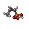


| #2: Chemical | ChemComp-FCN / Fosfomycin Mass: 138.059 Da / Num. of mol.: 1 / Source method: obtained synthetically / Formula: C3H7O4P / Comment: antibiotic*YM Mass: 138.059 Da / Num. of mol.: 1 / Source method: obtained synthetically / Formula: C3H7O4P / Comment: antibiotic*YM | ||||||||
|---|---|---|---|---|---|---|---|---|---|
| #3: Chemical |  Mass: 54.938 Da / Num. of mol.: 2 / Source method: obtained synthetically / Formula: Mn Mass: 54.938 Da / Num. of mol.: 2 / Source method: obtained synthetically / Formula: Mn#4: Chemical | ChemComp-NA /  Mass: 22.990 Da / Num. of mol.: 10 / Source method: obtained synthetically / Formula: Na Mass: 22.990 Da / Num. of mol.: 10 / Source method: obtained synthetically / Formula: Na#5: Chemical |  Mass: 39.098 Da / Num. of mol.: 2 / Source method: obtained synthetically / Formula: K Mass: 39.098 Da / Num. of mol.: 2 / Source method: obtained synthetically / Formula: K#6: Chemical | ChemComp-TLA / | Tartaric acid Mass: 150.087 Da / Num. of mol.: 1 / Source method: obtained synthetically / Formula: C4H6O6 / Feature type: SUBJECT OF INVESTIGATION Mass: 150.087 Da / Num. of mol.: 1 / Source method: obtained synthetically / Formula: C4H6O6 / Feature type: SUBJECT OF INVESTIGATION#7: Water | ChemComp-HOH / | WaterMass: 18.015 Da / Num. of mol.: 382 / Source method: isolated from a natural source / Formula: H2O |
-Details
| Has ligand of interest | Y |
|---|
-Experimental details
-Experiment
| Experiment | Method: X-RAY DIFFRACTION / Number of used crystals: 1 |
|---|
- Sample preparation
| Crystal | Density Matthews: 2.16 Å3/Da / Density % sol: 43.05 % |
|---|---|
| Crystal grow | Temperature: 289 K / Method: vapor diffusion, hanging drop Details: PEG3350 18% w/v, potassium sodium tartrate 0.2 M, MnCl2 6 mM |
-Data collection
| Diffraction | Mean temperature: 100 K / Serial crystal experiment: N |
|---|---|
| Diffraction source | Source: SYNCHROTRON / Site: MAX IV  / Beamline: BioMAX / Wavelength: 0.9919 Å / Beamline: BioMAX / Wavelength: 0.9919 Å |
| Detector | Type: DECTRIS EIGER X 16M / Detector: PIXEL / Date: May 29, 2019 |
| Radiation | Protocol: SINGLE WAVELENGTH / Monochromatic (M) / Laue (L): M / Scattering type: x-ray |
| Radiation wavelength | Wavelength: 0.9919 Å / Relative weight: 1 |
| Reflection | Resolution: 1.48→149.2 Å / Num. obs: 48410 / % possible obs: 99.4 % / Redundancy: 9.8 % / Biso Wilson estimate: 21.01 Å2 / CC1/2: 0.999 / Rrim(I) all: 0.089 / Net I/σ(I): 11.8 |
| Reflection shell | Resolution: 1.48→1.51 Å / Mean I/σ(I) obs: 1.2 / Num. unique obs: 2174 / CC1/2: 0.666 / Rrim(I) all: 1.452 / % possible all: 92.2 |
- Processing
| Software |
| |||||||||||||||||||||||||||||||||||||||||||||||||||||||||||||||||||||||||||||||||||||||||||||||||||||||||||||||||||||||
|---|---|---|---|---|---|---|---|---|---|---|---|---|---|---|---|---|---|---|---|---|---|---|---|---|---|---|---|---|---|---|---|---|---|---|---|---|---|---|---|---|---|---|---|---|---|---|---|---|---|---|---|---|---|---|---|---|---|---|---|---|---|---|---|---|---|---|---|---|---|---|---|---|---|---|---|---|---|---|---|---|---|---|---|---|---|---|---|---|---|---|---|---|---|---|---|---|---|---|---|---|---|---|---|---|---|---|---|---|---|---|---|---|---|---|---|---|---|---|---|---|
| Refinement | Method to determine structure: MOLECULAR REPLACEMENT / Resolution: 1.48→44.85 Å / SU ML: 0.1838 / Cross valid method: FREE R-VALUE / σ(F): 1.33 / Phase error: 24.4427 Stereochemistry target values: GeoStd + Monomer Library + CDL v1.2
| |||||||||||||||||||||||||||||||||||||||||||||||||||||||||||||||||||||||||||||||||||||||||||||||||||||||||||||||||||||||
| Solvent computation | Shrinkage radii: 0.9 Å / VDW probe radii: 1.1 Å / Solvent model: FLAT BULK SOLVENT MODEL | |||||||||||||||||||||||||||||||||||||||||||||||||||||||||||||||||||||||||||||||||||||||||||||||||||||||||||||||||||||||
| Displacement parameters | Biso mean: 27.59 Å2 | |||||||||||||||||||||||||||||||||||||||||||||||||||||||||||||||||||||||||||||||||||||||||||||||||||||||||||||||||||||||
| Refinement step | Cycle: LAST / Resolution: 1.48→44.85 Å
| |||||||||||||||||||||||||||||||||||||||||||||||||||||||||||||||||||||||||||||||||||||||||||||||||||||||||||||||||||||||
| Refine LS restraints |
| |||||||||||||||||||||||||||||||||||||||||||||||||||||||||||||||||||||||||||||||||||||||||||||||||||||||||||||||||||||||
| LS refinement shell |
|
