 Movie
Movie Controller

[English] 日本語
 Yorodumi
- PDB-8r02: Crystal structure of the retromer complex VPS29/VPS35 with the li... -
+ Open data

- Basic information
| Entry | Database: PDB / ID: 8r02 | ||||||
|---|---|---|---|---|---|---|---|
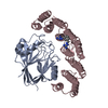
| Title | Crystal structure of the retromer complex VPS29/VPS35 with the ligand bis-1,3-phenyl guanylhydrazone, 2a | ||||||
 Components Components |
| ||||||
 Keywords Keywords |  PROTEIN TRANSPORT / Complex / transport / recycling / ligand PROTEIN TRANSPORT / Complex / transport / recycling / ligand | ||||||
| Function / homology |  Function and homology information Function and homology informationpositive regulation of locomotion involved in locomotory behavior / neurotransmitter receptor transport, endosome to plasma membrane / regulation of postsynapse assembly / mitochondrion-derived vesicle / negative regulation of protein localization / negative regulation of protein homooligomerization / regulation of dendritic spine maintenance / tubular endosome / mitochondrion to lysosome vesicle-mediated transport / positive regulation of Wnt protein secretion ...positive regulation of locomotion involved in locomotory behavior / neurotransmitter receptor transport, endosome to plasma membrane / regulation of postsynapse assembly / mitochondrion-derived vesicle / negative regulation of protein localization / negative regulation of protein homooligomerization / regulation of dendritic spine maintenance / tubular endosome / mitochondrion to lysosome vesicle-mediated transport / positive regulation of Wnt protein secretion / regulation of terminal button organization / retromer, cargo-selective complex / vesicle-mediated transport in synapse / WNT ligand biogenesis and trafficking / negative regulation of late endosome to lysosome transport / negative regulation of lysosomal protein catabolic process / positive regulation of dopamine receptor signaling pathway / positive regulation of dopamine biosynthetic process / mitochondrial fragmentation involved in apoptotic process / retromer complex / protein localization to endosome / dopaminergic synapse / regulation of synapse maturation / neurotransmitter receptor transport, endosome to postsynaptic membrane / voluntary musculoskeletal movement / regulation of protein metabolic process / transcytosis / endocytic recycling / positive regulation of protein localization to cell periphery / retrograde transport, endosome to Golgi / regulation of mitochondrion organization / positive regulation of mitochondrial fission / lysosome organization / regulation of presynapse assembly / D1 dopamine receptor binding / regulation of macroautophagy / intracellular protein transport / protein destabilization / modulation of chemical synaptic transmission / regulation of protein stability / Wnt signaling pathway / negative regulation of inflammatory response / positive regulation of protein catabolic process / positive regulation of canonical Wnt signaling pathway / late endosome / presynapse / postsynaptic density / lysosome / early endosome / endosome membrane / endosome / neuron projection / lysosomal membrane / negative regulation of gene expression / intracellular membrane-bounded organelle / neuronal cell body / glutamatergic synapse / positive regulation of gene expression / perinuclear region of cytoplasm / extracellular exosome / metal ion binding / cytosolSimilarity search - Function | ||||||
| Biological species |  Homo sapiens (human) Homo sapiens (human) | ||||||
| Method | X-RAY DIFFRACTION / SYNCHROTRON / MOLECULAR REPLACEMENT / Resolution: 2.5 Å | ||||||
 Authors Authors | Milani, M. / Fagnani, E. | ||||||
| Funding support |  Italy, 1items Italy, 1items
| ||||||
 Citation Citation | Journal: Comput Struct Biotechnol J / Year: 2024 Title: Stabilization of the retromer complex: Analysis of novel binding sites of bis-1,3-phenyl guanylhydrazone 2a to the VPS29/VPS35 interface. Authors: Fagnani, E. / Boni, F. / Seneci, P. / Gornati, D. / Muzio, L. / Mastrangelo, E. / Milani, M. | ||||||
| History |
|
- Structure visualization
| Structure viewer | Molecule: MolmilJmol/JSmol |
|---|
- Downloads & links
-Download
| PDBx/mmCIF format | 8r02.cif.gz | 515.6 KB | Display | PDBx/mmCIF format |
|---|---|---|---|---|
| PDB format | pdb8r02.ent.gz | 330.9 KB | Display | PDB format |
| PDBx/mmJSON format | 8r02.json.gz | Tree view | PDBx/mmJSON format | |
| Others |  Other downloads Other downloads |
-Validation report
| Arichive directory | https://data.pdbj.org/pub/pdb/validation_reports/r0/8r02ftp://data.pdbj.org/pub/pdb/validation_reports/r0/8r02 | HTTPS FTP |
|---|
-Related structure data
-Links
 PDBj
PDBj
- Assembly
| Deposited unit | 
| ||||||||||||||||||||||||||||||||||||||||||||||
|---|---|---|---|---|---|---|---|---|---|---|---|---|---|---|---|---|---|---|---|---|---|---|---|---|---|---|---|---|---|---|---|---|---|---|---|---|---|---|---|---|---|---|---|---|---|---|---|
| 1 | 
| ||||||||||||||||||||||||||||||||||||||||||||||
| 2 | 
| ||||||||||||||||||||||||||||||||||||||||||||||
| Unit cell |
| ||||||||||||||||||||||||||||||||||||||||||||||
| Noncrystallographic symmetry (NCS) | NCS domain:
NCS domain segments:
NCS ensembles :
|
-Components
| #1: Protein | Vacuole Mass: 20930.162 Da / Num. of mol.: 2 Source method: isolated from a genetically manipulated source Source: (gene. exp.) Homo sapiens (human) / Gene: VPS29 / Production host:  Escherichia coli (E. coli) / References: UniProt: Q9UBQ0 Escherichia coli (E. coli) / References: UniProt: Q9UBQ0#2: Protein | VacuoleMass: 35243.082 Da / Num. of mol.: 2 Source method: isolated from a genetically manipulated source Source: (gene. exp.) Homo sapiens (human) / Gene: VPS35 / Production host: Escherichia coli (E. coli) / References: UniProt: Q96QK1#3: Chemical | ChemComp-XFZ / | Mass: 246.272 Da / Num. of mol.: 1 / Source method: obtained synthetically / Formula: C10H14N8 / Feature type: SUBJECT OF INVESTIGATION #4: Water | ChemComp-HOH / | Water Mass: 18.015 Da / Num. of mol.: 64 / Source method: isolated from a natural source / Formula: H2O Mass: 18.015 Da / Num. of mol.: 64 / Source method: isolated from a natural source / Formula: H2OHas ligand of interest | Y | |
|---|
-Experimental details
-Experiment
| Experiment | Method: X-RAY DIFFRACTION / Number of used crystals: 1 |
|---|
- Sample preparation
| Crystal | Density Matthews: 2.58 Å3/Da / Density % sol: 52.37 % |
|---|---|
| Crystal grow | Temperature: 293 K / Method: vapor diffusion, sitting drop / pH: 7.4 Details: 20% PEG 3350, 150 mM NaK tartrate, 100 mM NaCl, pH 7.4 |
-Data collection
| Diffraction | Mean temperature: 100 K / Serial crystal experiment: N |
|---|---|
| Diffraction source | Source: SYNCHROTRON / Site: Diamond  / Beamline: I04-1 / Wavelength: 0.91788 Å / Beamline: I04-1 / Wavelength: 0.91788 Å |
| Detector | Type: DECTRIS PILATUS 6M-F / Detector: PIXEL / Date: Dec 12, 2021 |
| Radiation | Protocol: SINGLE WAVELENGTH / Monochromatic (M) / Laue (L): M / Scattering type: x-ray |
| Radiation wavelength | Wavelength: 0.91788 Å / Relative weight: 1 |
| Reflection | Resolution: 2.5→19.919 Å / Num. obs: 40494 / % possible obs: 99.7 % / Redundancy: 13.6 % / CC1/2: 0.999 / Net I/σ(I): 16.1 |
| Reflection shell | Resolution: 2.5→2.55 Å / Num. unique obs: 2913 / CC1/2: 0.597 |
- Processing
| Software |
| |||||||||||||||||||||||||||||||||||||||||||||||||||||||||||||||||||||||||||||||||||||||||||||||||||||||||||||||||||||||||||||||||||||||||||||||||||||||||||||||||||||||||||||||||||||||||||||||||||||||||||||||||||||||||||||||||||||||
|---|---|---|---|---|---|---|---|---|---|---|---|---|---|---|---|---|---|---|---|---|---|---|---|---|---|---|---|---|---|---|---|---|---|---|---|---|---|---|---|---|---|---|---|---|---|---|---|---|---|---|---|---|---|---|---|---|---|---|---|---|---|---|---|---|---|---|---|---|---|---|---|---|---|---|---|---|---|---|---|---|---|---|---|---|---|---|---|---|---|---|---|---|---|---|---|---|---|---|---|---|---|---|---|---|---|---|---|---|---|---|---|---|---|---|---|---|---|---|---|---|---|---|---|---|---|---|---|---|---|---|---|---|---|---|---|---|---|---|---|---|---|---|---|---|---|---|---|---|---|---|---|---|---|---|---|---|---|---|---|---|---|---|---|---|---|---|---|---|---|---|---|---|---|---|---|---|---|---|---|---|---|---|---|---|---|---|---|---|---|---|---|---|---|---|---|---|---|---|---|---|---|---|---|---|---|---|---|---|---|---|---|---|---|---|---|---|---|---|---|---|---|---|---|---|---|---|---|---|---|---|---|---|
| Refinement | Method to determine structure: MOLECULAR REPLACEMENT / Resolution: 2.5→19.919 Å / Cor.coef. Fo:Fc: 0.969 / Cor.coef. Fo:Fc free: 0.938 / WRfactor Rfree: 0.256 / WRfactor Rwork: 0.196 / SU B: 33.081 / SU ML: 0.323 / Average fsc free: 0.9279 / Average fsc work: 0.9448 / Cross valid method: FREE R-VALUE / ESU R: 0.537 / ESU R Free: 0.303 Details: Hydrogens have been added in their riding positions
| |||||||||||||||||||||||||||||||||||||||||||||||||||||||||||||||||||||||||||||||||||||||||||||||||||||||||||||||||||||||||||||||||||||||||||||||||||||||||||||||||||||||||||||||||||||||||||||||||||||||||||||||||||||||||||||||||||||||
| Solvent computation | Ion probe radii: 0.8 Å / Shrinkage radii: 0.8 Å / VDW probe radii: 1.2 Å / Solvent model: MASK BULK SOLVENT | |||||||||||||||||||||||||||||||||||||||||||||||||||||||||||||||||||||||||||||||||||||||||||||||||||||||||||||||||||||||||||||||||||||||||||||||||||||||||||||||||||||||||||||||||||||||||||||||||||||||||||||||||||||||||||||||||||||||
| Displacement parameters | Biso mean: 115.59 Å2
| |||||||||||||||||||||||||||||||||||||||||||||||||||||||||||||||||||||||||||||||||||||||||||||||||||||||||||||||||||||||||||||||||||||||||||||||||||||||||||||||||||||||||||||||||||||||||||||||||||||||||||||||||||||||||||||||||||||||
| Refinement step | Cycle: LAST / Resolution: 2.5→19.919 Å
| |||||||||||||||||||||||||||||||||||||||||||||||||||||||||||||||||||||||||||||||||||||||||||||||||||||||||||||||||||||||||||||||||||||||||||||||||||||||||||||||||||||||||||||||||||||||||||||||||||||||||||||||||||||||||||||||||||||||
| Refine LS restraints |
| |||||||||||||||||||||||||||||||||||||||||||||||||||||||||||||||||||||||||||||||||||||||||||||||||||||||||||||||||||||||||||||||||||||||||||||||||||||||||||||||||||||||||||||||||||||||||||||||||||||||||||||||||||||||||||||||||||||||
| Refine LS restraints NCS |
| |||||||||||||||||||||||||||||||||||||||||||||||||||||||||||||||||||||||||||||||||||||||||||||||||||||||||||||||||||||||||||||||||||||||||||||||||||||||||||||||||||||||||||||||||||||||||||||||||||||||||||||||||||||||||||||||||||||||
| LS refinement shell | Refine-ID: X-RAY DIFFRACTION / Total num. of bins used: 20
| |||||||||||||||||||||||||||||||||||||||||||||||||||||||||||||||||||||||||||||||||||||||||||||||||||||||||||||||||||||||||||||||||||||||||||||||||||||||||||||||||||||||||||||||||||||||||||||||||||||||||||||||||||||||||||||||||||||||
| Refinement TLS params. | Method: refined / Refine-ID: X-RAY DIFFRACTION
| |||||||||||||||||||||||||||||||||||||||||||||||||||||||||||||||||||||||||||||||||||||||||||||||||||||||||||||||||||||||||||||||||||||||||||||||||||||||||||||||||||||||||||||||||||||||||||||||||||||||||||||||||||||||||||||||||||||||
| Refinement TLS group | Selection: ALL |
