 Movie
Movie Controller


+ Open data

- Basic information
| Entry | Database: PDB / ID: 8qua | ||||||
|---|---|---|---|---|---|---|---|
| Title | GTP binding protein YsxC from Staphylococcus aureus | ||||||
 Components Components | Probable GTP-binding protein EngB | ||||||
 Keywords Keywords |  PROTEIN BINDING / YsxC / Ribosome / GTPase PROTEIN BINDING / YsxC / Ribosome / GTPase | ||||||
| Function / homology |  Function and homology information Function and homology information | ||||||
| Biological species |  Staphylococcus aureus subsp. aureus N315 (bacteria) Staphylococcus aureus subsp. aureus N315 (bacteria) | ||||||
| Method | X-RAY DIFFRACTION / MOLECULAR REPLACEMENT / Resolution: 2 Å | ||||||
 Authors Authors | Biktimirov, A. / Islamov, D. / Lazarenko, V. / Fatkhullin, B. / Validov, S. / Yusupov, M. / Usachev, K. | ||||||
| Funding support |  Russian Federation, 1items Russian Federation, 1items
| ||||||
 Citation Citation | Journal: Biochem.Biophys.Res.Commun. / Year: 2024 Title: Crystal structure of GTPase YsxC from Staphylococcus aureus. Authors: Biktimirov, A. / Islamov, D. / Fatkhullin, B. / Lazarenko, V. / Validov, S. / Yusupov, M. / Usachev, K. | ||||||
| History |
|
- Structure visualization
| Structure viewer | Molecule: MolmilJmol/JSmol |
|---|
- Downloads & links
-Download
| PDBx/mmCIF format | 8qua.cif.gz | 87.8 KB | Display | PDBx/mmCIF format |
|---|---|---|---|---|
| PDB format | pdb8qua.ent.gz | 64.8 KB | Display | PDB format |
| PDBx/mmJSON format | 8qua.json.gz | Tree view | PDBx/mmJSON format | |
| Others |  Other downloads Other downloads |
-Validation report
| Arichive directory | https://data.pdbj.org/pub/pdb/validation_reports/qu/8quaftp://data.pdbj.org/pub/pdb/validation_reports/qu/8qua | HTTPS FTP |
|---|
-Related structure data
| Similar structure data |
|---|
-Links
 PDBj
PDBj
- Assembly
| Deposited unit | 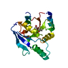
| ||||||||
|---|---|---|---|---|---|---|---|---|---|
| 1 |
| ||||||||
| Unit cell |
|
-Components
| #1: Protein | Mass: 22712.959 Da / Num. of mol.: 1 Source method: isolated from a genetically manipulated source Source: (gene. exp.) Staphylococcus aureus subsp. aureus N315 (bacteria)Gene: engB Production host: Escherichia coli 'BL21-Gold(DE3)pLysS AG' (bacteria)References: UniProt: A5ITJ8 |
|---|---|
| #2: Chemical | ChemComp-ACE / Acetyl group  Mass: 44.053 Da / Num. of mol.: 1 / Source method: obtained synthetically / Formula: C2H4O Mass: 44.053 Da / Num. of mol.: 1 / Source method: obtained synthetically / Formula: C2H4O |
| #3: Chemical | ChemComp-GOL / Glycerol  Mass: 92.094 Da / Num. of mol.: 1 / Source method: obtained synthetically / Formula: C3H8O3 Mass: 92.094 Da / Num. of mol.: 1 / Source method: obtained synthetically / Formula: C3H8O3 |
| #4: Water | ChemComp-HOH / Water Mass: 18.015 Da / Num. of mol.: 32 / Source method: isolated from a natural source / Formula: H2O Mass: 18.015 Da / Num. of mol.: 32 / Source method: isolated from a natural source / Formula: H2O |
| Has ligand of interest | N |
-Experimental details
-Experiment
| Experiment | Method: X-RAY DIFFRACTION / Number of used crystals: 1 |
|---|
- Sample preparation
| Crystal | Density Matthews: 1.94 Å3/Da / Density % sol: 36.74 % |
|---|---|
| Crystal grow | Temperature: 295.15 K / Method: vapor diffusion, hanging drop / pH: 7.2 / Details: 2.6M Potassium/sodium phosphate pH 7.6 / PH range: 6.8 - 7.4 |
-Data collection
| Diffraction | Mean temperature: 80 K / Serial crystal experiment: N | |||||||||||||||||||||
|---|---|---|---|---|---|---|---|---|---|---|---|---|---|---|---|---|---|---|---|---|---|---|
| Diffraction source | Source: SEALED TUBE / Type: RIGAKU PhotonJet-S / Wavelength: 1.54184 Å | |||||||||||||||||||||
| Detector | Type: RIGAKU HyPix-6000HE / Detector: PIXEL / Date: Aug 8, 2022 | |||||||||||||||||||||
| Radiation | Protocol: SINGLE WAVELENGTH / Monochromatic (M) / Laue (L): M / Scattering type: x-ray | |||||||||||||||||||||
| Radiation wavelength | Wavelength: 1.54184 Å / Relative weight: 1 | |||||||||||||||||||||
| Reflection | Resolution: 2→20.05 Å / Num. obs: 13613 / % possible obs: 99.6 % / Redundancy: 7.7 % / CC1/2: 0.998 / Rmerge(I) obs: 0.087 / Rpim(I) all: 0.044 / Rrim(I) all: 0.098 / Net I/σ(I): 11.3 | |||||||||||||||||||||
| Reflection shell | Diffraction-ID: 1
|
- Processing
| Software |
| |||||||||||||||||||||||||||||||||||||||||||||||||||||||||||||||||||||||||||||||||||||||||||||||||||||||||||||||||||||||||||||||||||||||||||||||||||||||||||||||||||||||||||||||||||||||||||||||||||||||||||||||||||||||||||||||||||||||
|---|---|---|---|---|---|---|---|---|---|---|---|---|---|---|---|---|---|---|---|---|---|---|---|---|---|---|---|---|---|---|---|---|---|---|---|---|---|---|---|---|---|---|---|---|---|---|---|---|---|---|---|---|---|---|---|---|---|---|---|---|---|---|---|---|---|---|---|---|---|---|---|---|---|---|---|---|---|---|---|---|---|---|---|---|---|---|---|---|---|---|---|---|---|---|---|---|---|---|---|---|---|---|---|---|---|---|---|---|---|---|---|---|---|---|---|---|---|---|---|---|---|---|---|---|---|---|---|---|---|---|---|---|---|---|---|---|---|---|---|---|---|---|---|---|---|---|---|---|---|---|---|---|---|---|---|---|---|---|---|---|---|---|---|---|---|---|---|---|---|---|---|---|---|---|---|---|---|---|---|---|---|---|---|---|---|---|---|---|---|---|---|---|---|---|---|---|---|---|---|---|---|---|---|---|---|---|---|---|---|---|---|---|---|---|---|---|---|---|---|---|---|---|---|---|---|---|---|---|---|---|---|---|
| Refinement | Method to determine structure: MOLECULAR REPLACEMENT / Resolution: 2→20.046 Å / Cor.coef. Fo:Fc: 0.918 / Cor.coef. Fo:Fc free: 0.886 / WRfactor Rfree: 0.303 / WRfactor Rwork: 0.245 / SU B: 5.75 / SU ML: 0.164 / Average fsc free: 0.9355 / Average fsc work: 0.9575 / Cross valid method: FREE R-VALUE / ESU R: 0.053 / ESU R Free: 0.047 Details: Hydrogens have been added in their riding positions
| |||||||||||||||||||||||||||||||||||||||||||||||||||||||||||||||||||||||||||||||||||||||||||||||||||||||||||||||||||||||||||||||||||||||||||||||||||||||||||||||||||||||||||||||||||||||||||||||||||||||||||||||||||||||||||||||||||||||
| Solvent computation | Ion probe radii: 0.8 Å / Shrinkage radii: 0.8 Å / VDW probe radii: 1.2 Å / Solvent model: MASK BULK SOLVENT | |||||||||||||||||||||||||||||||||||||||||||||||||||||||||||||||||||||||||||||||||||||||||||||||||||||||||||||||||||||||||||||||||||||||||||||||||||||||||||||||||||||||||||||||||||||||||||||||||||||||||||||||||||||||||||||||||||||||
| Displacement parameters | Biso mean: 37.306 Å2
| |||||||||||||||||||||||||||||||||||||||||||||||||||||||||||||||||||||||||||||||||||||||||||||||||||||||||||||||||||||||||||||||||||||||||||||||||||||||||||||||||||||||||||||||||||||||||||||||||||||||||||||||||||||||||||||||||||||||
| Refinement step | Cycle: LAST / Resolution: 2→20.046 Å
| |||||||||||||||||||||||||||||||||||||||||||||||||||||||||||||||||||||||||||||||||||||||||||||||||||||||||||||||||||||||||||||||||||||||||||||||||||||||||||||||||||||||||||||||||||||||||||||||||||||||||||||||||||||||||||||||||||||||
| Refine LS restraints |
| |||||||||||||||||||||||||||||||||||||||||||||||||||||||||||||||||||||||||||||||||||||||||||||||||||||||||||||||||||||||||||||||||||||||||||||||||||||||||||||||||||||||||||||||||||||||||||||||||||||||||||||||||||||||||||||||||||||||
| LS refinement shell | Refine-ID: X-RAY DIFFRACTION / Total num. of bins used: 20
|
