 Movie
Movie Controller


+ Open data

- Basic information
| Entry | Database: PDB / ID: 8qky | ||||||
|---|---|---|---|---|---|---|---|
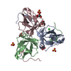
| Title | Bacteriophage T5 dUTPase | ||||||
 Components Components | Deoxyuridine 5'-triphosphate nucleotidohydrolase | ||||||
 Keywords Keywords |  HYDROLASE / bacteriophage / T5 / dUTPase / Deoxyuridine triphosphate nucleotidohydrolases / dUTP HYDROLASE / bacteriophage / T5 / dUTPase / Deoxyuridine triphosphate nucleotidohydrolases / dUTP | ||||||
| Function / homology |  Function and homology information Function and homology informationdUTP catabolic process / dUMP biosynthetic process / dUTP diphosphatase / dUTP diphosphatase activity / magnesium ion bindingSimilarity search - Function | ||||||
| Biological species |  Escherichia phage T5 (virus) Escherichia phage T5 (virus) | ||||||
| Method | X-RAY DIFFRACTION / SYNCHROTRON / MOLECULAR REPLACEMENT / Resolution: 2 Å | ||||||
 Authors Authors | Gabdulkhakov, A.G. / Dzhus, U.F. / Selikhanov, G.K. / Glukhov, A.S. | ||||||
| Funding support |  Russian Federation, 1items Russian Federation, 1items
| ||||||
 Citation Citation | Journal: Int J Mol Sci / Year: 2024 Title: Bacteriophage T5 dUTPase: Combination of Common Enzymatic and Novel Functions. Authors: Glukhov, A. / Marchenkov, V. / Dzhus, U. / Krutilina, A. / Selikhanov, G. / Gabdulkhakov, A. | ||||||
| History |
|
- Structure visualization
| Structure viewer | Molecule: MolmilJmol/JSmol |
|---|
- Downloads & links
-Download
| PDBx/mmCIF format | 8qky.cif.gz | 103.2 KB | Display | PDBx/mmCIF format |
|---|---|---|---|---|
| PDB format | pdb8qky.ent.gz | 71.2 KB | Display | PDB format |
| PDBx/mmJSON format | 8qky.json.gz | Tree view | PDBx/mmJSON format | |
| Others |  Other downloads Other downloads |
-Validation report
| Arichive directory | https://data.pdbj.org/pub/pdb/validation_reports/qk/8qkyftp://data.pdbj.org/pub/pdb/validation_reports/qk/8qky | HTTPS FTP |
|---|
-Related structure data
-Links
 PDBj
PDBj
- Assembly
| Deposited unit | 
| ||||||||||||
|---|---|---|---|---|---|---|---|---|---|---|---|---|---|
| 1 |
| ||||||||||||
| Unit cell |
|
-Components
| #1: Protein | Mass: 16223.650 Da / Num. of mol.: 3 Source method: isolated from a genetically manipulated source Source: (gene. exp.) Escherichia phage T5 (virus) / Gene: DUT / Production host:  Escherichia coli (E. coli) / References: UniProt: O48500 Escherichia coli (E. coli) / References: UniProt: O48500#2: Chemical | ChemComp-GOL / | Glycerol  Mass: 92.094 Da / Num. of mol.: 1 / Source method: obtained synthetically / Formula: C3H8O3 Mass: 92.094 Da / Num. of mol.: 1 / Source method: obtained synthetically / Formula: C3H8O3#3: Water | ChemComp-HOH / | Water Mass: 18.015 Da / Num. of mol.: 163 / Source method: isolated from a natural source / Formula: H2O Mass: 18.015 Da / Num. of mol.: 163 / Source method: isolated from a natural source / Formula: H2OHas ligand of interest | N | |
|---|
-Experimental details
-Experiment
| Experiment | Method: X-RAY DIFFRACTION / Number of used crystals: 1 |
|---|
- Sample preparation
| Crystal | Density Matthews: 2.75 Å3/Da / Density % sol: 55.22 % |
|---|---|
| Crystal grow | Temperature: 296 K / Method: vapor diffusion, sitting drop / pH: 8.5 Details: 0.075 M TRIS hydrochloride, 1.5 M Ammonium sulfate, 25% v/v glycerol |
-Data collection
| Diffraction | Mean temperature: 100 K / Serial crystal experiment: N |
|---|---|
| Diffraction source | Source: SYNCHROTRON / Site: PETRA III, DESY  / Beamline: P11 / Wavelength: 1.0332 Å / Beamline: P11 / Wavelength: 1.0332 Å |
| Detector | Type: DECTRIS PILATUS 6M / Detector: PIXEL / Date: Jun 15, 2019 |
| Radiation | Protocol: SINGLE WAVELENGTH / Monochromatic (M) / Laue (L): M / Scattering type: x-ray |
| Radiation wavelength | Wavelength: 1.0332 Å / Relative weight: 1 |
| Reflection | Resolution: 2→50 Å / Num. obs: 35488 / % possible obs: 99.9 % / Redundancy: 12.76 % / Biso Wilson estimate: 35.18 Å2 / CC1/2: 0.99 / Rmerge(I) obs: 0.07 / Net I/σ(I): 22.91 |
| Reflection shell | Resolution: 2→2.05 Å / Rmerge(I) obs: 1.01 / Mean I/σ(I) obs: 1.86 / Num. unique obs: 2637 / CC1/2: 0.71 |
- Processing
| Software |
| ||||||||||||||||||||||||||||||||||||||||||||||||||||||||||||||||||||||||||||||||||||||||||||||||||
|---|---|---|---|---|---|---|---|---|---|---|---|---|---|---|---|---|---|---|---|---|---|---|---|---|---|---|---|---|---|---|---|---|---|---|---|---|---|---|---|---|---|---|---|---|---|---|---|---|---|---|---|---|---|---|---|---|---|---|---|---|---|---|---|---|---|---|---|---|---|---|---|---|---|---|---|---|---|---|---|---|---|---|---|---|---|---|---|---|---|---|---|---|---|---|---|---|---|---|---|
| Refinement | Method to determine structure: MOLECULAR REPLACEMENT / Resolution: 2→46.76 Å / SU ML: 0.221 / Cross valid method: FREE R-VALUE / σ(F): 1.37 / Phase error: 23.5113 Stereochemistry target values: GeoStd + Monomer Library + CDL v1.2
| ||||||||||||||||||||||||||||||||||||||||||||||||||||||||||||||||||||||||||||||||||||||||||||||||||
| Solvent computation | Shrinkage radii: 0.9 Å / VDW probe radii: 1.11 Å / Solvent model: FLAT BULK SOLVENT MODEL | ||||||||||||||||||||||||||||||||||||||||||||||||||||||||||||||||||||||||||||||||||||||||||||||||||
| Displacement parameters | Biso mean: 40.21 Å2 | ||||||||||||||||||||||||||||||||||||||||||||||||||||||||||||||||||||||||||||||||||||||||||||||||||
| Refinement step | Cycle: LAST / Resolution: 2→46.76 Å
| ||||||||||||||||||||||||||||||||||||||||||||||||||||||||||||||||||||||||||||||||||||||||||||||||||
| Refine LS restraints |
| ||||||||||||||||||||||||||||||||||||||||||||||||||||||||||||||||||||||||||||||||||||||||||||||||||
| LS refinement shell |
|
