 Movie
Movie Controller

[English] 日本語
 Yorodumi
- PDB-8qi7: Cryo-EM Structure of Human Serine Hydroxymethyltransferase, isofo... -
+ Open data

- Basic information
| Entry | Database: PDB / ID: 8qi7 | ||||||
|---|---|---|---|---|---|---|---|
| Title | Cryo-EM Structure of Human Serine Hydroxymethyltransferase, isoform 2 (SHMT2) | ||||||
 Components Components | Serine hydroxymethyltransferase, mitochondrial | ||||||
 Keywords Keywords | TRANSFERASE / one-carbon metabolism / folate cycle / tetrahydtofolate / mitochondria | ||||||
| Function / homology |  Function and homology information Function and homology informationBRISC complex / L-allo-threonine aldolase activity / regulation of mitochondrial translation / purine nucleobase biosynthetic process / L-serine metabolic process / glycine metabolic process / L-serine biosynthetic process / serine binding / glycine hydroxymethyltransferase / glycine hydroxymethyltransferase activity ...BRISC complex / L-allo-threonine aldolase activity / regulation of mitochondrial translation / purine nucleobase biosynthetic process / L-serine metabolic process / glycine metabolic process / L-serine biosynthetic process / serine binding / glycine hydroxymethyltransferase / glycine hydroxymethyltransferase activity / glycine biosynthetic process from serine / L-serine catabolic process / Metabolism of folate and pterines / regulation of oxidative phosphorylation / tetrahydrofolate metabolic process / response to type I interferon / protein K63-linked deubiquitination / tetrahydrofolate interconversion / regulation of aerobic respiration / mitochondrial nucleoid / folic acid metabolic process / RHOG GTPase cycle / mRNA regulatory element binding translation repressor activity / protein tetramerization / mRNA 5'-UTR binding / microtubule cytoskeleton / pyridoxal phosphate binding / one-carbon metabolic process / protein homotetramerization / mitochondrial inner membrane / mitochondrial matrix / chromatin binding / positive regulation of cell population proliferation / protein homodimerization activity / mitochondrion / extracellular exosome / zinc ion binding / nucleus / cytosol / cytoplasmSimilarity search - Function | ||||||
| Biological species |  Homo sapiens (human) Homo sapiens (human) | ||||||
| Method | ELECTRON MICROSCOPY / single particle reconstruction / cryo EM / Resolution: 2.9 Å | ||||||
 Authors Authors | Rutkiewicz, M. / Tran, L.H. / Ruszkowski, M. | ||||||
| Funding support |  Poland, 1items Poland, 1items
| ||||||
 Citation Citation | Journal: To Be Published Title: New Structural Models of SHMT2 Authors: Rutkiewicz, M. / Tran, L.H. / Ruszkowski, M. #1: Journal: Acta Crystallogr D Struct Biol / Year: 2019 Title: Macromolecular structure determination using X-rays, neutrons and electrons: recent developments in Phenix. Authors: Dorothee Liebschner / Pavel V Afonine / Matthew L Baker / Gábor Bunkóczi / Vincent B Chen / Tristan I Croll / Bradley Hintze / Li Wei Hung / Swati Jain / Airlie J McCoy / Nigel W Moriarty ...Authors: Dorothee Liebschner / Pavel V Afonine / Matthew L Baker / Gábor Bunkóczi / Vincent B Chen / Tristan I Croll / Bradley Hintze / Li Wei Hung / Swati Jain / Airlie J McCoy / Nigel W Moriarty / Robert D Oeffner / Billy K Poon / Michael G Prisant / Randy J Read / Jane S Richardson / David C Richardson / Massimo D Sammito / Oleg V Sobolev / Duncan H Stockwell / Thomas C Terwilliger / Alexandre G Urzhumtsev / Lizbeth L Videau / Christopher J Williams / Paul D Adams /    Abstract: Diffraction (X-ray, neutron and electron) and electron cryo-microscopy are powerful methods to determine three-dimensional macromolecular structures, which are required to understand biological ...Diffraction (X-ray, neutron and electron) and electron cryo-microscopy are powerful methods to determine three-dimensional macromolecular structures, which are required to understand biological processes and to develop new therapeutics against diseases. The overall structure-solution workflow is similar for these techniques, but nuances exist because the properties of the reduced experimental data are different. Software tools for structure determination should therefore be tailored for each method. Phenix is a comprehensive software package for macromolecular structure determination that handles data from any of these techniques. Tasks performed with Phenix include data-quality assessment, map improvement, model building, the validation/rebuilding/refinement cycle and deposition. Each tool caters to the type of experimental data. The design of Phenix emphasizes the automation of procedures, where possible, to minimize repetitive and time-consuming manual tasks, while default parameters are chosen to encourage best practice. A graphical user interface provides access to many command-line features of Phenix and streamlines the transition between programs, project tracking and re-running of previous tasks. | ||||||
| History |
|
- Structure visualization
| Structure viewer | Molecule: MolmilJmol/JSmol |
|---|
- Downloads & links
-Download
| PDBx/mmCIF format | 8qi7.cif.gz | 394.6 KB | Display | PDBx/mmCIF format |
|---|---|---|---|---|
| PDB format | pdb8qi7.ent.gz | 292.2 KB | Display | PDB format |
| PDBx/mmJSON format | 8qi7.json.gz | Tree view | PDBx/mmJSON format | |
| Others |  Other downloads Other downloads |
-Validation report
| Arichive directory | https://data.pdbj.org/pub/pdb/validation_reports/qi/8qi7ftp://data.pdbj.org/pub/pdb/validation_reports/qi/8qi7 | HTTPS FTP |
|---|
-Related structure data
| Related structure data | 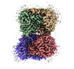 18436MC M: map data used to model this data C: citing same article ( |
|---|---|
| Similar structure data |

-Links
 PDBj
PDBj

- Assembly
| Deposited unit | 
| ||||||||||||||||||||||||||||||||||||||||||||||
|---|---|---|---|---|---|---|---|---|---|---|---|---|---|---|---|---|---|---|---|---|---|---|---|---|---|---|---|---|---|---|---|---|---|---|---|---|---|---|---|---|---|---|---|---|---|---|---|
| 1 |
| ||||||||||||||||||||||||||||||||||||||||||||||
| Noncrystallographic symmetry (NCS) | NCS domain:
NCS domain segments: Component-ID: 1 / Ens-ID: ens_1 / Beg auth comp-ID: TRP / Beg label comp-ID: TRP / End auth comp-ID: HIS / End label comp-ID: HIS / Auth seq-ID: 43 - 504 / Label seq-ID: 15 - 476
NCS oper:
|
-Components
| #1: Protein | / SHMT / Glycine hydroxymethyltransferase / Serine methylase Mass: 52944.973 Da / Num. of mol.: 4 Source method: isolated from a genetically manipulated source Source: (gene. exp.) Homo sapiens (human) / Gene: SHMT2 / Plasmid: pMCSG68Production host:  Escherichia coli 'BL21-Gold(DE3)pLysS AG' (bacteria) Escherichia coli 'BL21-Gold(DE3)pLysS AG' (bacteria)References: UniProt: P34897, glycine hydroxymethyltransferase#2: Water | ChemComp-HOH / | Water Mass: 18.015 Da / Num. of mol.: 117 / Source method: isolated from a natural source / Formula: H2O Mass: 18.015 Da / Num. of mol.: 117 / Source method: isolated from a natural source / Formula: H2OHas ligand of interest | N | |
|---|
-Experimental details
-Experiment
| Experiment | Method: ELECTRON MICROSCOPY |
|---|---|
| EM experiment | Aggregation state: PARTICLE / 3D reconstruction method: single particle reconstruction |
- Sample preparation
| Component | Name: SHMT2 in the form of PLP internal aldimineSerine hydroxymethyltransferase Type: COMPLEX / Entity ID: #1 / Source: RECOMBINANT |
|---|---|
| Source (natural) | Organism: Homo sapiens (human) |
| Source (recombinant) | Organism: Escherichia coli 'BL21-Gold(DE3)pLysS AG' (bacteria) |
| Buffer solution | pH: 7.5 / Details: 25 mM Hepes pH 7.5, 150 mM NaCl, 1 mM TCEP |
| Specimen | Conc.: 0.6 mg/ml / Embedding applied: NO / Shadowing applied: NO / Staining applied: NO / Vitrification applied: YES |
| Specimen support | Grid material: COPPER |
| Vitrification | Instrument: FEI VITROBOT MARK IV / Cryogen name: ETHANE / Humidity: 95 % / Chamber temperature: 277 K |
- Electron microscopy imaging
| Experimental equipment |  Model: Titan Krios / Image courtesy: FEI Company |
|---|---|
| Microscopy | Model: FEI TITAN KRIOS |
| Electron gun | Electron source: FIELD EMISSION GUN / Accelerating voltage: 300 kV / Illumination mode: FLOOD BEAM |
| Electron lens | Mode: BRIGHT FIELDBright-field microscopy / Nominal defocus max: 3000 nm / Nominal defocus min: 500 nm / Cs: 2.7 mm |
| Image recording | Electron dose: 40.4 e/Å2 / Film or detector model: GATAN K3 BIOQUANTUM (6k x 4k) / Num. of grids imaged: 1 / Num. of real images: 7948 |
| Image scans | Width: 5760 / Height: 4092 |
- Processing
| EM software |
| ||||||||||||||||||||||||||||||||||||||||
|---|---|---|---|---|---|---|---|---|---|---|---|---|---|---|---|---|---|---|---|---|---|---|---|---|---|---|---|---|---|---|---|---|---|---|---|---|---|---|---|---|---|
| CTF correction | Type: NONE | ||||||||||||||||||||||||||||||||||||||||
| Symmetry | Point symmetry: D2 (2x2 fold dihedral) | ||||||||||||||||||||||||||||||||||||||||
| 3D reconstruction | Resolution: 2.9 Å / Resolution method: FSC 0.143 CUT-OFF / Num. of particles: 146577 / Algorithm: FOURIER SPACE / Num. of class averages: 29 / Symmetry type: POINT | ||||||||||||||||||||||||||||||||||||||||
| Atomic model building | Protocol: FLEXIBLE FIT / Space: REAL | ||||||||||||||||||||||||||||||||||||||||
| Atomic model building | PDB-ID: 8aql Pdb chain-ID: A / Accession code: 8aql / Source name: PDB / Type: experimental model | ||||||||||||||||||||||||||||||||||||||||
| Refinement | Cross valid method: NONE Stereochemistry target values: GeoStd + Monomer Library + CDL v1.2 | ||||||||||||||||||||||||||||||||||||||||
| Displacement parameters | Biso mean: 63.35 Å2 | ||||||||||||||||||||||||||||||||||||||||
| Refine LS restraints |
| ||||||||||||||||||||||||||||||||||||||||
| Refine LS restraints NCS |
|
