 Movie
Movie Controller


+ Open data

- Basic information
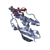
| Entry | Database: PDB / ID: 8p2a | ||||||
|---|---|---|---|---|---|---|---|
| Title | Crystal structure of SaFMN3 domain 2 | ||||||
 Components Components | FMN-binding domain-containing protein | ||||||
 Keywords Keywords |  FLAVOPROTEIN / multi-flavinylated protein / FMN / post-translational FLAVOPROTEIN / multi-flavinylated protein / FMN / post-translational | ||||||
| Function / homology | FMN-binding / FMN-binding domain / FMN_bind / Twin arginine translocation (Tat) signal profile. / Twin-arginine translocation pathway, signal sequence / FMN binding / membrane / FLAVIN MONONUCLEOTIDE / FMN-binding domain-containing protein Function and homology information Function and homology information | ||||||
| Biological species |  Streptomyces azureus (bacteria) Streptomyces azureus (bacteria) | ||||||
| Method | X-RAY DIFFRACTION / SYNCHROTRON / MOLECULAR REPLACEMENT / Resolution: 2 Å | ||||||
 Authors Authors | Rozeboom, H.J. / Fraaije, M.W. | ||||||
| Funding support | 1items
| ||||||
 Citation Citation | Journal: Bba Adv / Year: 2023 Title: Characterization of two bacterial multi-flavinylated proteins harboring multiple covalent flavin cofactors. Authors: Tong, Y. / Rozeboom, H.J. / Loonstra, M.R. / Wijma, H.J. / Fraaije, M.W. | ||||||
| History |
|
- Structure visualization
| Structure viewer | Molecule: MolmilJmol/JSmol |
|---|
- Downloads & links
-Download
| PDBx/mmCIF format | 8p2a.cif.gz | 48.3 KB | Display | PDBx/mmCIF format |
|---|---|---|---|---|
| PDB format | pdb8p2a.ent.gz | 33.4 KB | Display | PDB format |
| PDBx/mmJSON format | 8p2a.json.gz | Tree view | PDBx/mmJSON format | |
| Others |  Other downloads Other downloads |
-Validation report
| Arichive directory | https://data.pdbj.org/pub/pdb/validation_reports/p2/8p2aftp://data.pdbj.org/pub/pdb/validation_reports/p2/8p2a | HTTPS FTP |
|---|
-Related structure data
-Links
 PDBj
PDBj

- Assembly
| Deposited unit | 
| ||||||||
|---|---|---|---|---|---|---|---|---|---|
| 1 |
| ||||||||
| Unit cell |
| ||||||||
| Components on special symmetry positions |
|
-Components
| #1: Protein | Mass: 8945.968 Da / Num. of mol.: 1 Source method: isolated from a genetically manipulated source Source: (gene. exp.) Streptomyces azureus (bacteria) / Gene: SAZU_6878 / Production host: Escherichia coli (E. coli) / Strain (production host): NEB 10-beta / References: UniProt: A0A0K8PVQ4 |
|---|---|
| #2: Chemical | ChemComp-FMN / Flavin mononucleotide  Mass: 456.344 Da / Num. of mol.: 1 / Source method: isolated from a natural source / Formula: C17H21N4O9P / Feature type: SUBJECT OF INVESTIGATION Mass: 456.344 Da / Num. of mol.: 1 / Source method: isolated from a natural source / Formula: C17H21N4O9P / Feature type: SUBJECT OF INVESTIGATION |
| #3: Water | ChemComp-HOH / Water Mass: 18.015 Da / Num. of mol.: 23 / Source method: isolated from a natural source / Formula: H2O Mass: 18.015 Da / Num. of mol.: 23 / Source method: isolated from a natural source / Formula: H2O |
| Has ligand of interest | Y |
-Experimental details
-Experiment
| Experiment | Method: X-RAY DIFFRACTION / Number of used crystals: 1 |
|---|
- Sample preparation
| Crystal | Density Matthews: 1.9 Å3/Da / Density % sol: 35 % |
|---|---|
| Crystal grow | Temperature: 294 K / Method: vapor diffusion, sitting drop / Details: 38 - 42 % PEG3350, 0.05 M bis-tris / PH range: 6.5 - 7.0 |
-Data collection
| Diffraction | Mean temperature: 100 K / Serial crystal experiment: N |
|---|---|
| Diffraction source | Source: SYNCHROTRON / Site: ESRF  / Beamline: MASSIF-1 / Wavelength: 0.9655 Å / Beamline: MASSIF-1 / Wavelength: 0.9655 Å |
| Detector | Type: DECTRIS PILATUS3 6M / Detector: PIXEL / Date: Apr 22, 2022 |
| Radiation | Protocol: SINGLE WAVELENGTH / Monochromatic (M) / Laue (L): M / Scattering type: x-ray |
| Radiation wavelength | Wavelength: 0.9655 Å / Relative weight: 1 |
| Reflection | Resolution: 2→45.6 Å / Num. obs: 5999 / % possible obs: 100 % / Redundancy: 13.1 % / CC1/2: 0.996 / Rmerge(I) obs: 0.14 / Rpim(I) all: 0.039 / Rrim(I) all: 0.146 / Χ2: 0.79 / Net I/σ(I): 9 / Num. measured all: 78876 |
| Reflection shell | Resolution: 2→2.05 Å / % possible obs: 100 % / Redundancy: 11 % / Rmerge(I) obs: 0.452 / Num. measured all: 4505 / Num. unique obs: 408 / CC1/2: 0.982 / Rpim(I) all: 0.137 / Rrim(I) all: 0.473 / Χ2: 0.37 / Net I/σ(I) obs: 2.4 |
- Processing
| Software |
| ||||||||||||||||||||||||||||||||||||||||||||||||||||||||||||||||||||||||||||||||||||||||||||||||||||||||||||||||||||||||||||||||||||||||||||||||||||||||||||||||||||||||||||||||||||||
|---|---|---|---|---|---|---|---|---|---|---|---|---|---|---|---|---|---|---|---|---|---|---|---|---|---|---|---|---|---|---|---|---|---|---|---|---|---|---|---|---|---|---|---|---|---|---|---|---|---|---|---|---|---|---|---|---|---|---|---|---|---|---|---|---|---|---|---|---|---|---|---|---|---|---|---|---|---|---|---|---|---|---|---|---|---|---|---|---|---|---|---|---|---|---|---|---|---|---|---|---|---|---|---|---|---|---|---|---|---|---|---|---|---|---|---|---|---|---|---|---|---|---|---|---|---|---|---|---|---|---|---|---|---|---|---|---|---|---|---|---|---|---|---|---|---|---|---|---|---|---|---|---|---|---|---|---|---|---|---|---|---|---|---|---|---|---|---|---|---|---|---|---|---|---|---|---|---|---|---|---|---|---|---|
| Refinement | Method to determine structure: MOLECULAR REPLACEMENT / Resolution: 2→30.42 Å / Cor.coef. Fo:Fc: 0.948 / Cor.coef. Fo:Fc free: 0.915 / SU B: 14.4 / SU ML: 0.208 / Cross valid method: THROUGHOUT / ESU R: 0.228 / ESU R Free: 0.211 / Stereochemistry target values: MAXIMUM LIKELIHOOD / Details: HYDROGENS HAVE BEEN ADDED IN THE RIDING POSITIONS
| ||||||||||||||||||||||||||||||||||||||||||||||||||||||||||||||||||||||||||||||||||||||||||||||||||||||||||||||||||||||||||||||||||||||||||||||||||||||||||||||||||||||||||||||||||||||
| Solvent computation | Ion probe radii: 0.8 Å / Shrinkage radii: 0.8 Å / VDW probe radii: 1.2 Å / Solvent model: MASK | ||||||||||||||||||||||||||||||||||||||||||||||||||||||||||||||||||||||||||||||||||||||||||||||||||||||||||||||||||||||||||||||||||||||||||||||||||||||||||||||||||||||||||||||||||||||
| Displacement parameters | Biso mean: 46.793 Å2
| ||||||||||||||||||||||||||||||||||||||||||||||||||||||||||||||||||||||||||||||||||||||||||||||||||||||||||||||||||||||||||||||||||||||||||||||||||||||||||||||||||||||||||||||||||||||
| Refinement step | Cycle: 1 / Resolution: 2→30.42 Å
| ||||||||||||||||||||||||||||||||||||||||||||||||||||||||||||||||||||||||||||||||||||||||||||||||||||||||||||||||||||||||||||||||||||||||||||||||||||||||||||||||||||||||||||||||||||||
| Refine LS restraints |
|