 Movie
Movie Controller

[English] 日本語
 Yorodumi
- PDB-8p22: X-ray structure of acetylcholine-binding protein (AChBP) in compl... -
+ Open data

- Basic information
| Entry | Database: PDB / ID: 8p22 | ||||||
|---|---|---|---|---|---|---|---|
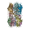
| Title | X-ray structure of acetylcholine-binding protein (AChBP) in complex with IOTA376. | ||||||
 Components Components | Acetylcholine-binding protein | ||||||
 Keywords Keywords | CHOLINE-BINDING PROTEIN / Fragment based drug design / Acetylcholine-binding protein / choline-binding proteins | ||||||
| Function / homology |  Function and homology information Function and homology information acetylcholine receptor activity / acetylcholine-gated monoatomic cation-selective channel activity / synaptic cleft / response to nicotine / neuron projection / synapse / membrane acetylcholine receptor activity / acetylcholine-gated monoatomic cation-selective channel activity / synaptic cleft / response to nicotine / neuron projection / synapse / membraneSimilarity search - Function | ||||||
| Biological species |   Lymnaea stagnalis (great pond snail) Lymnaea stagnalis (great pond snail) | ||||||
| Method | X-RAY DIFFRACTION / SYNCHROTRON / MOLECULAR REPLACEMENT / Resolution: 2.2 Å | ||||||
 Authors Authors | Cederfelt, D. / Boronat, P. / Dobritzsch, D. / Hennig, S. / Fitzgerald, E.A. / de Esch, I.J.P. / Danielson, U.H. | ||||||
| Funding support | European Union, 1items
| ||||||
 Citation Citation | Journal: To Be Published Title: Elucidating the regulation of ligand gated ion channels via biophysical studies of ligand-induced conformational dynamics of acetylcholine binding proteins Authors: Fitzgerald, E.A. / Cederfelt, D. / Boronat, P. / Aarmo Lund, B. / Dobritzsch, D. / de Esch, I.J.P. / Danielson, U.H. | ||||||
| History |
|
- Structure visualization
| Structure viewer | Molecule: MolmilJmol/JSmol |
|---|
- Downloads & links
-Download
| PDBx/mmCIF format | 8p22.cif.gz | 420.3 KB | Display | PDBx/mmCIF format |
|---|---|---|---|---|
| PDB format | pdb8p22.ent.gz | 344.7 KB | Display | PDB format |
| PDBx/mmJSON format | 8p22.json.gz | Tree view | PDBx/mmJSON format | |
| Others |  Other downloads Other downloads |
-Validation report
| Arichive directory | https://data.pdbj.org/pub/pdb/validation_reports/p2/8p22ftp://data.pdbj.org/pub/pdb/validation_reports/p2/8p22 | HTTPS FTP |
|---|
-Related structure data


-Links
 PDBj
PDBj

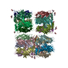
- Assembly
| Deposited unit | 
| ||||||||
|---|---|---|---|---|---|---|---|---|---|
| 1 |
| ||||||||
| Unit cell |
|
-Components
| #1: Protein | Mass: 23454.029 Da / Num. of mol.: 10 Source method: isolated from a genetically manipulated source Source: (gene. exp.) Lymnaea stagnalis (great pond snail) / Production host:   Spodoptera frugiperda (fall armyworm) / References: UniProt: P58154 Spodoptera frugiperda (fall armyworm) / References: UniProt: P58154#2: Chemical | ChemComp-WNO / Mass: 254.330 Da / Num. of mol.: 9 / Source method: obtained synthetically / Formula: C15H18N4 / Feature type: SUBJECT OF INVESTIGATION #3: Chemical | Sulfate  Mass: 96.063 Da / Num. of mol.: 3 / Source method: obtained synthetically / Formula: SO4 Mass: 96.063 Da / Num. of mol.: 3 / Source method: obtained synthetically / Formula: SO4#4: Chemical | ChemComp-GOL / | Glycerol  Mass: 92.094 Da / Num. of mol.: 1 / Source method: obtained synthetically / Formula: C3H8O3 Mass: 92.094 Da / Num. of mol.: 1 / Source method: obtained synthetically / Formula: C3H8O3#5: Water | ChemComp-HOH / | Water Mass: 18.015 Da / Num. of mol.: 825 / Source method: isolated from a natural source / Formula: H2O Mass: 18.015 Da / Num. of mol.: 825 / Source method: isolated from a natural source / Formula: H2OHas ligand of interest | Y | |
|---|
-Experimental details
-Experiment
| Experiment | Method: X-RAY DIFFRACTION / Number of used crystals: 1 |
|---|
- Sample preparation
| Crystal | Density Matthews: 2.13 Å3/Da / Density % sol: 42.32 % |
|---|---|
| Crystal grow | Temperature: 293 K / Method: vapor diffusion, hanging drop Details: PEG 3350 3% Ammonium sulphate 1.8M HEPES buffer 0.1M, pH 7.75 |
-Data collection
| Diffraction | Mean temperature: 100 K / Serial crystal experiment: N |
|---|---|
| Diffraction source | Source: SYNCHROTRON / Site: MAX IV  / Beamline: BioMAX / Wavelength: 0.9762 Å / Beamline: BioMAX / Wavelength: 0.9762 Å |
| Detector | Type: DECTRIS EIGER X 16M / Detector: PIXEL / Date: Apr 4, 2020 |
| Radiation | Protocol: MAD / Monochromatic (M) / Laue (L): M / Scattering type: x-ray |
| Radiation wavelength | Wavelength: 0.9762 Å / Relative weight: 1 |
| Reflection | Resolution: 1.53→999 Å / Num. obs: 101962 / % possible obs: 88.32 % / Redundancy: 2 % / CC1/2: 0.907 / Rrim(I) all: 0.1852 / Net I/σ(I): 7.41 |
| Reflection shell | Resolution: 2.2→2.279 Å / Num. unique obs: 10549 / CC1/2: 0.588 / Rrim(I) all: 0.8643 |
- Processing
| Software |
| ||||||||||||||||||||||||||||||||||||||||||||||||||||||||||||||||||||||||||||||||||||||||||||||||||||||||||||||||||||||||||||||||||||||||||||||||||||||||||||||||||||||||||||||||||||||
|---|---|---|---|---|---|---|---|---|---|---|---|---|---|---|---|---|---|---|---|---|---|---|---|---|---|---|---|---|---|---|---|---|---|---|---|---|---|---|---|---|---|---|---|---|---|---|---|---|---|---|---|---|---|---|---|---|---|---|---|---|---|---|---|---|---|---|---|---|---|---|---|---|---|---|---|---|---|---|---|---|---|---|---|---|---|---|---|---|---|---|---|---|---|---|---|---|---|---|---|---|---|---|---|---|---|---|---|---|---|---|---|---|---|---|---|---|---|---|---|---|---|---|---|---|---|---|---|---|---|---|---|---|---|---|---|---|---|---|---|---|---|---|---|---|---|---|---|---|---|---|---|---|---|---|---|---|---|---|---|---|---|---|---|---|---|---|---|---|---|---|---|---|---|---|---|---|---|---|---|---|---|---|---|
| Refinement | Method to determine structure: MOLECULAR REPLACEMENT / Resolution: 2.2→49.51 Å / Cor.coef. Fo:Fc: 0.945 / SU B: 11.666 / SU ML: 0.245 / Cross valid method: THROUGHOUT / ESU R: 0.376 / Stereochemistry target values: MAXIMUM LIKELIHOOD / Details: HYDROGENS HAVE BEEN ADDED IN THE RIDING POSITIONS
| ||||||||||||||||||||||||||||||||||||||||||||||||||||||||||||||||||||||||||||||||||||||||||||||||||||||||||||||||||||||||||||||||||||||||||||||||||||||||||||||||||||||||||||||||||||||
| Solvent computation | Ion probe radii: 0.8 Å / Shrinkage radii: 0.8 Å / VDW probe radii: 1.2 Å / Solvent model: MASK | ||||||||||||||||||||||||||||||||||||||||||||||||||||||||||||||||||||||||||||||||||||||||||||||||||||||||||||||||||||||||||||||||||||||||||||||||||||||||||||||||||||||||||||||||||||||
| Displacement parameters | Biso mean: 46.067 Å2
| ||||||||||||||||||||||||||||||||||||||||||||||||||||||||||||||||||||||||||||||||||||||||||||||||||||||||||||||||||||||||||||||||||||||||||||||||||||||||||||||||||||||||||||||||||||||
| Refinement step | Cycle: 1 / Resolution: 2.2→49.51 Å
| ||||||||||||||||||||||||||||||||||||||||||||||||||||||||||||||||||||||||||||||||||||||||||||||||||||||||||||||||||||||||||||||||||||||||||||||||||||||||||||||||||||||||||||||||||||||
| Refine LS restraints |
|