 Movie
Movie Controller


+ Open data

- Basic information
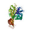
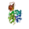
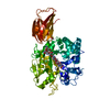
| Entry | Database: PDB / ID: 8oye | ||||||
|---|---|---|---|---|---|---|---|
| Title | Clostridium perfringens chitinase CP4_3455 E196Q with chitin | ||||||
 Components Components | Chitodextrinase | ||||||
 Keywords Keywords |  HYDROLASE / chitinase / TIM / substrate / necrotic enteritis HYDROLASE / chitinase / TIM / substrate / necrotic enteritis | ||||||
| Function / homology |  Function and homology informationpolysaccharide binding / chitin binding / hydrolase activity, hydrolyzing O-glycosyl compounds / carbohydrate metabolic process Function and homology informationpolysaccharide binding / chitin binding / hydrolase activity, hydrolyzing O-glycosyl compounds / carbohydrate metabolic processSimilarity search - Function | ||||||
| Biological species |   Clostridium perfringens (bacteria) Clostridium perfringens (bacteria) | ||||||
| Method | X-RAY DIFFRACTION / SYNCHROTRON / MOLECULAR REPLACEMENT / Resolution: 1.35 Å | ||||||
 Authors Authors | Bloch, Y. / Savvides, S.N. | ||||||
| Funding support |  Belgium, 1items Belgium, 1items
| ||||||
 Citation Citation | Journal: To Be Published Title: Clostridium perfringens chitinase CP4_3455 E196Q with chitin Authors: Bloch, Y. / Savvides, S.N. | ||||||
| History |
|
- Structure visualization
| Structure viewer | Molecule: MolmilJmol/JSmol |
|---|
- Downloads & links
-Download
| PDBx/mmCIF format | 8oye.cif.gz | 427.6 KB | Display | PDBx/mmCIF format |
|---|---|---|---|---|
| PDB format | pdb8oye.ent.gz | 295.1 KB | Display | PDB format |
| PDBx/mmJSON format | 8oye.json.gz | Tree view | PDBx/mmJSON format | |
| Others |  Other downloads Other downloads |
-Validation report
| Arichive directory | https://data.pdbj.org/pub/pdb/validation_reports/oy/8oyeftp://data.pdbj.org/pub/pdb/validation_reports/oy/8oye | HTTPS FTP |
|---|
-Related structure data
| Related structure data | |
|---|---|
| Similar structure data |


-Links
 PDBj
PDBj
- Assembly
| Deposited unit | 
| ||||||||||||
|---|---|---|---|---|---|---|---|---|---|---|---|---|---|
| 1 |
| ||||||||||||
| Unit cell |
|
-Components
| #1: Protein | Mass: 65171.520 Da / Num. of mol.: 1 / Mutation: E196Q active site mutant Source method: isolated from a genetically manipulated source Details: Residues [1-2] (MG) cloning scar / start codon. Truncated construct compared to WT. Residues [569-] (SHHHHHH) cloning scar and purification tag. Present during crystallization. Source: (gene. exp.) Clostridium perfringens (bacteria) / Gene: CP4_3455 / Plasmid: pET / Production host: Escherichia coli (E. coli) / Strain (production host): T7 Express lysY/Iq / References: UniProt: F8UNI5 |
|---|---|
| #2: Polysaccharide | 2-acetamido-2-deoxy-beta-D-glucopyranose-(1-4)-2-acetamido-2-deoxy-beta-D-glucopyranose-(1-4)-2- ...2-acetamido-2-deoxy-beta-D-glucopyranose-(1-4)-2-acetamido-2-deoxy-beta-D-glucopyranose-(1-4)-2-acetamido-2-deoxy-beta-D-glucopyranose-(1-4)-2-acetamido-2-deoxy-beta-D-glucopyranose-(1-4)-2-acetamido-2-deoxy-beta-D-glucopyranose / Mass: 1033.979 Da / Num. of mol.: 1 Source method: isolated from a genetically manipulated source |
| #3: Chemical | ChemComp-DMS / Dimethyl sulfoxide  Mass: 78.133 Da / Num. of mol.: 1 / Source method: obtained synthetically / Formula: C2H6OS / Comment: DMSO, precipitant*YM Mass: 78.133 Da / Num. of mol.: 1 / Source method: obtained synthetically / Formula: C2H6OS / Comment: DMSO, precipitant*YM |
| #4: Water | ChemComp-HOH / Water Mass: 18.015 Da / Num. of mol.: 576 / Source method: isolated from a natural source / Formula: H2O Mass: 18.015 Da / Num. of mol.: 576 / Source method: isolated from a natural source / Formula: H2O |
| Has ligand of interest | Y |
-Experimental details
-Experiment
| Experiment | Method: X-RAY DIFFRACTION / Number of used crystals: 1 |
|---|
- Sample preparation
| Crystal | Density Matthews: 2.35 Å3/Da / Density % sol: 47.75 % |
|---|---|
| Crystal grow | Temperature: 287 K / Method: vapor diffusion, sitting drop Details: PEGION A1: 0.2 M Sodium fluoride 20% w/v Polyethylene glycol 3,350, 1mM chitin hexamer (seeded) |
-Data collection
| Diffraction | Mean temperature: 100 K / Serial crystal experiment: N | ||||||||||||||||||||||||||||
|---|---|---|---|---|---|---|---|---|---|---|---|---|---|---|---|---|---|---|---|---|---|---|---|---|---|---|---|---|---|
| Diffraction source | Source: SYNCHROTRON / Site: PETRA III, EMBL c/o DESY  / Beamline: P14 (MX2) / Wavelength: 0.9763 Å / Beamline: P14 (MX2) / Wavelength: 0.9763 Å | ||||||||||||||||||||||||||||
| Detector | Type: DECTRIS EIGER X 16M / Detector: PIXEL / Date: Aug 25, 2019 | ||||||||||||||||||||||||||||
| Radiation | Protocol: SINGLE WAVELENGTH / Monochromatic (M) / Laue (L): M / Scattering type: x-ray | ||||||||||||||||||||||||||||
| Radiation wavelength | Wavelength: 0.9763 Å / Relative weight: 1 | ||||||||||||||||||||||||||||
| Reflection | Resolution: 1.35→77.95 Å / Num. obs: 134505 / % possible obs: 99.3 % / Redundancy: 12.01 % / Biso Wilson estimate: 17.66 Å2 / CC1/2: 0.999 / Rrim(I) all: 0.058 / Net I/σ(I): 23.82 | ||||||||||||||||||||||||||||
| Reflection shell | Diffraction-ID: 1
|
- Processing
| Software |
| |||||||||||||||||||||||||||||||||||||||||||||||||||||||||||||||||||||||||||||||||||||||||||||||||||||||||
|---|---|---|---|---|---|---|---|---|---|---|---|---|---|---|---|---|---|---|---|---|---|---|---|---|---|---|---|---|---|---|---|---|---|---|---|---|---|---|---|---|---|---|---|---|---|---|---|---|---|---|---|---|---|---|---|---|---|---|---|---|---|---|---|---|---|---|---|---|---|---|---|---|---|---|---|---|---|---|---|---|---|---|---|---|---|---|---|---|---|---|---|---|---|---|---|---|---|---|---|---|---|---|---|---|---|---|
| Refinement | Method to determine structure: MOLECULAR REPLACEMENT / Resolution: 1.35→77.95 Å / SU ML: 0.1176 / Cross valid method: FREE R-VALUE / σ(F): 1.34 / Phase error: 15.4072 Stereochemistry target values: GeoStd + Monomer Library + CDL v1.2
| |||||||||||||||||||||||||||||||||||||||||||||||||||||||||||||||||||||||||||||||||||||||||||||||||||||||||
| Solvent computation | Shrinkage radii: 0.9 Å / VDW probe radii: 1.11 Å / Solvent model: FLAT BULK SOLVENT MODEL | |||||||||||||||||||||||||||||||||||||||||||||||||||||||||||||||||||||||||||||||||||||||||||||||||||||||||
| Displacement parameters | Biso mean: 26.27 Å2 | |||||||||||||||||||||||||||||||||||||||||||||||||||||||||||||||||||||||||||||||||||||||||||||||||||||||||
| Refinement step | Cycle: LAST / Resolution: 1.35→77.95 Å
| |||||||||||||||||||||||||||||||||||||||||||||||||||||||||||||||||||||||||||||||||||||||||||||||||||||||||
| Refine LS restraints |
| |||||||||||||||||||||||||||||||||||||||||||||||||||||||||||||||||||||||||||||||||||||||||||||||||||||||||
| LS refinement shell |
|
