 Movie
Movie Controller

[English] 日本語
 Yorodumi
- PDB-8oql: Structure of Mycobacterium tuberculosis beta-oxidation trifunctio... -
+ Open data

- Basic information
| Entry | Database: PDB / ID: 8oql | ||||||||||||
|---|---|---|---|---|---|---|---|---|---|---|---|---|---|
| Title | Structure of Mycobacterium tuberculosis beta-oxidation trifunctional enzyme in complex with Fragment-M-1 | ||||||||||||
 Components Components |
| ||||||||||||
 Keywords Keywords |  OXIDOREDUCTASE / fatty acid beta oxidation complex / mycobacterium tuberculosis / TFE / fragment screening / substrate channeling OXIDOREDUCTASE / fatty acid beta oxidation complex / mycobacterium tuberculosis / TFE / fragment screening / substrate channeling | ||||||||||||
| Function / homology |  Function and homology informationlong-chain-3-hydroxyacyl-CoA dehydrogenase activity / enoyl-CoA hydratase activity / fatty acid beta-oxidation / acyltransferase activity, transferring groups other than amino-acyl groups / NAD+ binding / Transferases; Acyltransferases; Transferring groups other than aminoacyl groups / peptidoglycan-based cell wall / plasma membrane / cytosol Function and homology informationlong-chain-3-hydroxyacyl-CoA dehydrogenase activity / enoyl-CoA hydratase activity / fatty acid beta-oxidation / acyltransferase activity, transferring groups other than amino-acyl groups / NAD+ binding / Transferases; Acyltransferases; Transferring groups other than aminoacyl groups / peptidoglycan-based cell wall / plasma membrane / cytosolSimilarity search - Function | ||||||||||||
| Biological species |  Mycobacterium tuberculosis H37Rv (bacteria) Mycobacterium tuberculosis H37Rv (bacteria) | ||||||||||||
| Method | X-RAY DIFFRACTION / SYNCHROTRON / MOLECULAR REPLACEMENT / Resolution: 2.7 Å | ||||||||||||
 Authors Authors | Dalwani, S. / Wierenga, R.K. / Venkatesan, R. | ||||||||||||
| Funding support |  Finland, 3items Finland, 3items
| ||||||||||||
 Citation Citation | Journal: Biorxiv / Year: 2024 Title: Crystallographic fragment binding studies of the Mycobacterium tuberculosis trifunctional enzyme suggest binding pockets for the tails of the acyl-CoA substrates at its active sites and a ...Title: Crystallographic fragment binding studies of the Mycobacterium tuberculosis trifunctional enzyme suggest binding pockets for the tails of the acyl-CoA substrates at its active sites and a potential substrate channeling path between them Authors: Dalwani, S. / Metz, A. / Huschmann, F.U. / Weiss, M.S. / Wierenga, R.K. / Venkatesan, R. #1: Journal: J Struct Biol / Year: 2021Title: Substrate specificity and conformational flexibility properties of the Mycobacterium tuberculosis beta-oxidation trifunctional enzyme. Authors: Dalwani, S. #2: Journal: ACS Chem Biol / Year: 2013Title: Structure of mycobacterial beta-oxidation trifunctional enzyme reveals its altered assembly and putative substrate channeling pathway. Authors: Venkatesan, R. / Wierenga, R.K. | ||||||||||||
| History |
|
- Structure visualization
| Structure viewer | Molecule: MolmilJmol/JSmol |
|---|
- Downloads & links
-Download
| PDBx/mmCIF format | 8oql.cif.gz | 1013.7 KB | Display | PDBx/mmCIF format |
|---|---|---|---|---|
| PDB format | pdb8oql.ent.gz | 702.3 KB | Display | PDB format |
| PDBx/mmJSON format | 8oql.json.gz | Tree view | PDBx/mmJSON format | |
| Others |  Other downloads Other downloads |
-Validation report
| Arichive directory | https://data.pdbj.org/pub/pdb/validation_reports/oq/8oqlftp://data.pdbj.org/pub/pdb/validation_reports/oq/8oql | HTTPS FTP |
|---|
-Related structure data
| Related structure data | 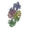 8opuC 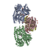 8opvC  8opwC 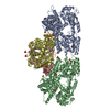 8opxC  8opyC  8oqmC  8oqnC 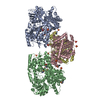 8oqoC  8oqpC 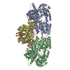 8oqqC 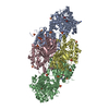 8oqrC 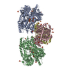 8oqsC 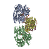 8oqtC  8oquC 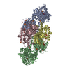 8oqvC C: citing same article ( |
|---|---|
| Similar structure data |
-Links
 PDBj
PDBj


- Assembly
| Deposited unit | 
| ||||||||||||
|---|---|---|---|---|---|---|---|---|---|---|---|---|---|
| 1 |
| ||||||||||||
| Unit cell |
|
-Components
-Protein , 2 types, 4 molecules ABCD
| #1: Protein | Mass: 78005.805 Da / Num. of mol.: 2 Source method: isolated from a genetically manipulated source Source: (gene. exp.) Mycobacterium tuberculosis H37Rv (bacteria)Gene: fadB, Rv0860 / Production host: Escherichia coli BL21(DE3) (bacteria)References: UniProt: O53872, 3-hydroxyacyl-CoA dehydrogenase#2: Protein | Mass: 42476.355 Da / Num. of mol.: 2 Source method: isolated from a genetically manipulated source Source: (gene. exp.) Mycobacterium tuberculosis H37Rv (bacteria)Gene: fadA, Rv0859 / Production host: Escherichia coli BL21(DE3) (bacteria)References: UniProt: O53871, Transferases; Acyltransferases; Transferring groups other than aminoacyl groups |
|---|
-Non-polymers , 5 types, 146 molecules 


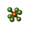

| #3: Chemical | ChemComp-SO4 / Sulfate Mass: 96.063 Da / Num. of mol.: 34 / Source method: obtained synthetically / Formula: SO4 Mass: 96.063 Da / Num. of mol.: 34 / Source method: obtained synthetically / Formula: SO4#4: Chemical | ChemComp-GOL / Glycerol Mass: 92.094 Da / Num. of mol.: 4 / Source method: obtained synthetically / Formula: C3H8O3 Mass: 92.094 Da / Num. of mol.: 4 / Source method: obtained synthetically / Formula: C3H8O3#5: Chemical | ChemComp-ARF / Formamide Type: L-peptide NH3 amino terminus / Mass: 45.041 Da / Num. of mol.: 6 / Source method: obtained synthetically / Formula: CH3NO Type: L-peptide NH3 amino terminus / Mass: 45.041 Da / Num. of mol.: 6 / Source method: obtained synthetically / Formula: CH3NO#6: Chemical | ChemComp-A9J /  Mass: 144.964 Da / Num. of mol.: 25 / Source method: obtained synthetically / Formula: F6P / Feature type: SUBJECT OF INVESTIGATION Mass: 144.964 Da / Num. of mol.: 25 / Source method: obtained synthetically / Formula: F6P / Feature type: SUBJECT OF INVESTIGATION#7: Water | ChemComp-HOH / | WaterMass: 18.015 Da / Num. of mol.: 77 / Source method: isolated from a natural source / Formula: H2O |
|---|
-Details
| Has ligand of interest | Y |
|---|
-Experimental details
-Experiment
| Experiment | Method: X-RAY DIFFRACTION / Number of used crystals: 1 |
|---|
- Sample preparation
| Crystal | Density Matthews: 3.89 Å3/Da / Density % sol: 68.37 % |
|---|---|
| Crystal grow | Temperature: 295 K / Method: vapor diffusion, hanging drop / pH: 8.5 / Details: 2 M Ammonium Sulfate, 0.1M Tris pH 8.5 |
-Data collection
| Diffraction | Mean temperature: 100 K / Serial crystal experiment: N |
|---|---|
| Diffraction source | Source: SYNCHROTRON / Site: MAX IV  / Beamline: BioMAX / Wavelength: 0.968619 Å / Beamline: BioMAX / Wavelength: 0.968619 Å |
| Detector | Type: DECTRIS EIGER X 16M / Detector: PIXEL / Date: Jun 7, 2019 |
| Radiation | Protocol: SINGLE WAVELENGTH / Monochromatic (M) / Laue (L): M / Scattering type: x-ray |
| Radiation wavelength | Wavelength: 0.968619 Å / Relative weight: 1 |
| Reflection | Resolution: 2.7→48.32 Å / Num. obs: 100954 / % possible obs: 99.7 % / Redundancy: 4.8 % / Biso Wilson estimate: 71.56 Å2 / CC1/2: 0.998 / Net I/σ(I): 10.9 |
| Reflection shell | Resolution: 2.7→2.75 Å / Mean I/σ(I) obs: 1.3 / Num. unique obs: 4803 / CC1/2: 0.538 |
- Processing
| Software |
| |||||||||||||||||||||||||||||||||||||||||||||||||||||||||||||||||||||||||||||||||||||||||||||||||||||||||||||||||||||||||||||||||||||||||||||||||||||||||||||||||||||||||||||||||||||||||||||||||||||||||||||||||||||||||
|---|---|---|---|---|---|---|---|---|---|---|---|---|---|---|---|---|---|---|---|---|---|---|---|---|---|---|---|---|---|---|---|---|---|---|---|---|---|---|---|---|---|---|---|---|---|---|---|---|---|---|---|---|---|---|---|---|---|---|---|---|---|---|---|---|---|---|---|---|---|---|---|---|---|---|---|---|---|---|---|---|---|---|---|---|---|---|---|---|---|---|---|---|---|---|---|---|---|---|---|---|---|---|---|---|---|---|---|---|---|---|---|---|---|---|---|---|---|---|---|---|---|---|---|---|---|---|---|---|---|---|---|---|---|---|---|---|---|---|---|---|---|---|---|---|---|---|---|---|---|---|---|---|---|---|---|---|---|---|---|---|---|---|---|---|---|---|---|---|---|---|---|---|---|---|---|---|---|---|---|---|---|---|---|---|---|---|---|---|---|---|---|---|---|---|---|---|---|---|---|---|---|---|---|---|---|---|---|---|---|---|---|---|---|---|---|---|---|---|
| Refinement | Method to determine structure: MOLECULAR REPLACEMENT / Resolution: 2.7→48.32 Å / SU ML: 0.3726 / Cross valid method: FREE R-VALUE / σ(F): 0.87 / Phase error: 24.2557 Stereochemistry target values: GeoStd + Monomer Library + CDL v1.2
| |||||||||||||||||||||||||||||||||||||||||||||||||||||||||||||||||||||||||||||||||||||||||||||||||||||||||||||||||||||||||||||||||||||||||||||||||||||||||||||||||||||||||||||||||||||||||||||||||||||||||||||||||||||||||
| Solvent computation | Shrinkage radii: 0.9 Å / VDW probe radii: 1.11 Å / Solvent model: FLAT BULK SOLVENT MODEL | |||||||||||||||||||||||||||||||||||||||||||||||||||||||||||||||||||||||||||||||||||||||||||||||||||||||||||||||||||||||||||||||||||||||||||||||||||||||||||||||||||||||||||||||||||||||||||||||||||||||||||||||||||||||||
| Displacement parameters | Biso mean: 74.02 Å2 | |||||||||||||||||||||||||||||||||||||||||||||||||||||||||||||||||||||||||||||||||||||||||||||||||||||||||||||||||||||||||||||||||||||||||||||||||||||||||||||||||||||||||||||||||||||||||||||||||||||||||||||||||||||||||
| Refinement step | Cycle: LAST / Resolution: 2.7→48.32 Å
| |||||||||||||||||||||||||||||||||||||||||||||||||||||||||||||||||||||||||||||||||||||||||||||||||||||||||||||||||||||||||||||||||||||||||||||||||||||||||||||||||||||||||||||||||||||||||||||||||||||||||||||||||||||||||
| Refine LS restraints |
| |||||||||||||||||||||||||||||||||||||||||||||||||||||||||||||||||||||||||||||||||||||||||||||||||||||||||||||||||||||||||||||||||||||||||||||||||||||||||||||||||||||||||||||||||||||||||||||||||||||||||||||||||||||||||
| LS refinement shell |
| |||||||||||||||||||||||||||||||||||||||||||||||||||||||||||||||||||||||||||||||||||||||||||||||||||||||||||||||||||||||||||||||||||||||||||||||||||||||||||||||||||||||||||||||||||||||||||||||||||||||||||||||||||||||||
| Refinement TLS params. | Method: refined / Origin x: -43.8407768051 Å / Origin y: 34.1416952805 Å / Origin z: -1.64715510787 Å
| |||||||||||||||||||||||||||||||||||||||||||||||||||||||||||||||||||||||||||||||||||||||||||||||||||||||||||||||||||||||||||||||||||||||||||||||||||||||||||||||||||||||||||||||||||||||||||||||||||||||||||||||||||||||||
| Refinement TLS group | Selection details: all |
