 Movie
Movie Controller

[English] 日本語
 Yorodumi
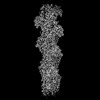
- PDB-8oid: Cryo-EM structure of ADP-bound, filamentous beta-actin harboring ... -
+ Open data

- Basic information
| Entry | Database: PDB / ID: 8oid | ||||||||||||
|---|---|---|---|---|---|---|---|---|---|---|---|---|---|
| Title | Cryo-EM structure of ADP-bound, filamentous beta-actin harboring the N111S mutation | ||||||||||||
 Components Components | Actin, cytoplasmic 1, N-terminally processed | ||||||||||||
 Keywords Keywords | STRUCTURAL PROTEIN / Actin filament / cytoskeletal protein / ATPase | ||||||||||||
| Function / homology |  Function and homology information Function and homology informationpositive regulation of norepinephrine uptake / cellular response to cytochalasin B / bBAF complex / npBAF complex / postsynaptic actin cytoskeleton organization / regulation of transepithelial transport / brahma complex / nBAF complex / structural constituent of postsynaptic actin cytoskeleton / morphogenesis of a polarized epithelium ...positive regulation of norepinephrine uptake / cellular response to cytochalasin B / bBAF complex / npBAF complex / postsynaptic actin cytoskeleton organization / regulation of transepithelial transport / brahma complex / nBAF complex / structural constituent of postsynaptic actin cytoskeleton / morphogenesis of a polarized epithelium / Formation of annular gap junctions / GBAF complex / Gap junction degradation / postsynaptic actin cytoskeleton / protein localization to adherens junction / regulation of G0 to G1 transition / dense body / Cell-extracellular matrix interactions / Tat protein binding / Folding of actin by CCT/TriC / regulation of double-strand break repair / regulation of nucleotide-excision repair / RSC-type complex / apical protein localization / Prefoldin mediated transfer of substrate to CCT/TriC / adherens junction assembly / RHOF GTPase cycle / Adherens junctions interactions / tight junction / Sensory processing of sound by outer hair cells of the cochlea / SWI/SNF complex / Interaction between L1 and Ankyrins / Sensory processing of sound by inner hair cells of the cochlea / regulation of mitotic metaphase/anaphase transition / regulation of norepinephrine uptake / positive regulation of double-strand break repair / positive regulation of T cell differentiation / NuA4 histone acetyltransferase complex / regulation of synaptic vesicle endocytosis / apical junction complex / maintenance of blood-brain barrier / establishment or maintenance of cell polarity / cortical cytoskeleton / positive regulation of double-strand break repair via homologous recombination / positive regulation of stem cell population maintenance / nitric-oxide synthase binding / Recycling pathway of L1 / regulation of cyclin-dependent protein serine/threonine kinase activity / regulation of G1/S transition of mitotic cell cycle / negative regulation of cell differentiation / brush border / kinesin binding / calyx of Held / EPH-ephrin mediated repulsion of cells / RHO GTPases Activate WASPs and WAVEs / RHO GTPases activate IQGAPs / positive regulation of myoblast differentiation / regulation of protein localization to plasma membrane / EPHB-mediated forward signaling / substantia nigra development / axonogenesis / negative regulation of protein binding / actin filament / cell motility / RHO GTPases Activate Formins / Translocation of SLC2A4 (GLUT4) to the plasma membrane / regulation of transmembrane transporter activity / positive regulation of cell differentiation / FCGR3A-mediated phagocytosis / adherens junction / Hydrolases; Acting on acid anhydrides; Acting on acid anhydrides to facilitate cellular and subcellular movement / DNA Damage Recognition in GG-NER / tau protein binding / Signaling by high-kinase activity BRAF mutants / Schaffer collateral - CA1 synapse / MAP2K and MAPK activation / B-WICH complex positively regulates rRNA expression / structural constituent of cytoskeleton / cytoplasmic ribonucleoprotein granule / kinetochore / Regulation of actin dynamics for phagocytic cup formation / platelet aggregation / nuclear matrix / VEGFA-VEGFR2 Pathway / UCH proteinases / Signaling by RAF1 mutants / Signaling by moderate kinase activity BRAF mutants / Paradoxical activation of RAF signaling by kinase inactive BRAF / Signaling downstream of RAS mutants / nucleosome / cell-cell junction / Signaling by BRAF and RAF1 fusions / actin cytoskeleton / presynapse / lamellipodium / Clathrin-mediated endocytosis / Factors involved in megakaryocyte development and platelet production / HATs acetylate histones / blood microparticle / regulation of apoptotic processSimilarity search - Function | ||||||||||||
| Biological species |  Homo sapiens (human) Homo sapiens (human) | ||||||||||||
| Method | ELECTRON MICROSCOPY / single particle reconstruction / cryo EM / Resolution: 2.3 Å | ||||||||||||
 Authors Authors | Oosterheert, W. / Blanc, F.E.C. / Roy, A. / Belyy, A. / Hofnagel, O. / Hummer, G. / Bieling, P. / Raunser, S. | ||||||||||||
| Funding support |  Germany, European Union, 3items Germany, European Union, 3items
| ||||||||||||
 Citation Citation | Journal: Nat Struct Mol Biol / Year: 2023 Title: Molecular mechanisms of inorganic-phosphate release from the core and barbed end of actin filaments. Authors: Wout Oosterheert / Florian E C Blanc / Ankit Roy / Alexander Belyy / Micaela Boiero Sanders / Oliver Hofnagel / Gerhard Hummer / Peter Bieling / Stefan Raunser / Abstract: The release of inorganic phosphate (P) from actin filaments constitutes a key step in their regulated turnover, which is fundamental to many cellular functions. The mechanisms underlying P release ...The release of inorganic phosphate (P) from actin filaments constitutes a key step in their regulated turnover, which is fundamental to many cellular functions. The mechanisms underlying P release from the core and barbed end of actin filaments remain unclear. Here, using human and bovine actin isoforms, we combine cryo-EM with molecular-dynamics simulations and in vitro reconstitution to demonstrate how actin releases P through a 'molecular backdoor'. While constantly open at the barbed end, the backdoor is predominantly closed in filament-core subunits and opens only transiently through concerted amino acid rearrangements. This explains why P escapes rapidly from the filament end but slowly from internal subunits. In a nemaline-myopathy-associated actin variant, the backdoor is predominantly open in filament-core subunits, resulting in accelerated P release and filaments with drastically shortened ADP-P caps. Our results provide the molecular basis for P release from actin and exemplify how a disease-linked mutation distorts the nucleotide-state distribution and atomic structure of the filament. | ||||||||||||
| History |
|
- Structure visualization
| Structure viewer | Molecule: MolmilJmol/JSmol |
|---|
- Downloads & links
-Download
| PDBx/mmCIF format | 8oid.cif.gz | 405.7 KB | Display | PDBx/mmCIF format |
|---|---|---|---|---|
| PDB format | pdb8oid.ent.gz | 277.5 KB | Display | PDB format |
| PDBx/mmJSON format | 8oid.json.gz | Tree view | PDBx/mmJSON format | |
| Others |  Other downloads Other downloads |
-Validation report
| Arichive directory | https://data.pdbj.org/pub/pdb/validation_reports/oi/8oidftp://data.pdbj.org/pub/pdb/validation_reports/oi/8oid | HTTPS FTP |
|---|
-Related structure data
| Related structure data | 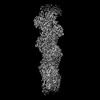 16889MC 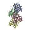 8oi6C 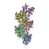 8oi8C C: citing same article ( M: map data used to model this data |
|---|---|
| Similar structure data |
-Links
 PDBj
PDBj


- Assembly
| Deposited unit | 
| ||||||||||||||||||||||||||||||||||||||||||||||||||||||||||||||||||||||||||||||||||||||||||||||||||||||||||||||||||||||||||||||||||||||
|---|---|---|---|---|---|---|---|---|---|---|---|---|---|---|---|---|---|---|---|---|---|---|---|---|---|---|---|---|---|---|---|---|---|---|---|---|---|---|---|---|---|---|---|---|---|---|---|---|---|---|---|---|---|---|---|---|---|---|---|---|---|---|---|---|---|---|---|---|---|---|---|---|---|---|---|---|---|---|---|---|---|---|---|---|---|---|---|---|---|---|---|---|---|---|---|---|---|---|---|---|---|---|---|---|---|---|---|---|---|---|---|---|---|---|---|---|---|---|---|---|---|---|---|---|---|---|---|---|---|---|---|---|---|---|---|
| 1 |
| ||||||||||||||||||||||||||||||||||||||||||||||||||||||||||||||||||||||||||||||||||||||||||||||||||||||||||||||||||||||||||||||||||||||
| Noncrystallographic symmetry (NCS) | NCS domain:
NCS domain segments: Ens-ID: ens_1
|