Method to determine structure: MOLECULAR REPLACEMENT / Resolution: 3.1→55.87 Å / Cor.coef. Fo:Fc: 0.927 / Cor.coef. Fo:Fc free: 0.868 / SU B: 115.018 / SU ML: 0.849 / Cross valid method: THROUGHOUT / ESU R Free: 0.719 / Stereochemistry target values: MAXIMUM LIKELIHOOD / Details: HYDROGENS HAVE BEEN ADDED IN THE RIDING POSITIONS
Rfactor
Num. reflection
% reflection
Selection details
Rfree
0.33672
1004
4.9 %
RANDOM
Rwork
0.234
-
-
-
obs
0.23903
19502
99.89 %
-
Solvent computation
Ion probe radii: 0.8 Å / Shrinkage radii: 0.8 Å / VDW probe radii: 1.2 Å / Solvent model: MASK
 Movie
Movie Controller


 Open data
 Basic information
 Components
 Keywords
 Function and homology information
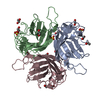
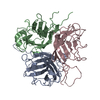
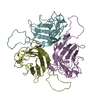
 Human adenovirus 24
 Authors
 United Kingdom, 2items
  Citation
 Structure visualization
 Downloads & links
 Other downloads


 PDBj
PDBj
 Assembly
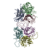

 Sample preparation
 Processing