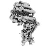
 Movie
Movie Controller

[English] 日本語
 Yorodumi
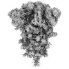
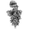
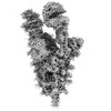
- PDB-8jyk: Structure of the SARS-CoV-2 XBB.1.5 spike glycoprotein (closed st... -
+ Open data

- Basic information
| Entry | Database: PDB / ID: 8jyk | |||||||||||||||||||||
|---|---|---|---|---|---|---|---|---|---|---|---|---|---|---|---|---|---|---|---|---|---|---|
| Title | Structure of the SARS-CoV-2 XBB.1.5 spike glycoprotein (closed state 1) | |||||||||||||||||||||
 Components Components | Spike glycoprotein Spike protein Spike protein | |||||||||||||||||||||
 Keywords Keywords | VIRAL PROTEIN / spike protein / glycoprotein / VIRUS | |||||||||||||||||||||
| Function / homology |  Function and homology information Function and homology informationMaturation of spike protein / viral translation / Translation of Structural Proteins / Virion Assembly and Release / host cell surface / host extracellular space / suppression by virus of host tetherin activity / Induction of Cell-Cell Fusion / structural constituent of virion / host cell endoplasmic reticulum-Golgi intermediate compartment membrane ...Maturation of spike protein / viral translation / Translation of Structural Proteins / Virion Assembly and Release / host cell surface / host extracellular space / suppression by virus of host tetherin activity / Induction of Cell-Cell Fusion / structural constituent of virion / host cell endoplasmic reticulum-Golgi intermediate compartment membrane / entry receptor-mediated virion attachment to host cell / receptor-mediated endocytosis of virus by host cell / Attachment and Entry / membrane fusion / positive regulation of viral entry into host cell / receptor-mediated virion attachment to host cell / receptor ligand activity / host cell surface receptor binding / fusion of virus membrane with host plasma membrane / fusion of virus membrane with host endosome membrane / viral envelope / symbiont-mediated suppression of host type I interferon-mediated signaling pathway / virion attachment to host cell / SARS-CoV-2 activates/modulates innate and adaptive immune responses / host cell plasma membrane / virion membrane / membrane / identical protein binding / plasma membraneSimilarity search - Function | |||||||||||||||||||||
| Biological species |   Severe acute respiratory syndrome coronavirus 2 Severe acute respiratory syndrome coronavirus 2 | |||||||||||||||||||||
| Method | ELECTRON MICROSCOPY / single particle reconstruction / cryo EM / Resolution: 2.59 Å | |||||||||||||||||||||
 Authors Authors | Yajima, H. / Anraku, Y. / Kita, S. / Kimura, K. / Sasaki, J. / Sasaki-Tabata, K. / Maenaka, K. / Hashiguchi, T. | |||||||||||||||||||||
| Funding support |  Japan, 6items Japan, 6items
| |||||||||||||||||||||
 Citation Citation | Journal: To Be Published Title: Virological characteristics of the SARS-CoV-2 XBB.1.5 variant Authors: Tamura, T. / Ito, J. / Uriu, K. / Zahradnik, J. / Kida, I. / Anraku, Y. / Nasser, H. / Shofa, M. / Oda, Y. / Lytras, S. / Nao, N. / Itakura, Y. / Deguchi, S. / Suzuki, R. / Wang, L. / Begum, ...Authors: Tamura, T. / Ito, J. / Uriu, K. / Zahradnik, J. / Kida, I. / Anraku, Y. / Nasser, H. / Shofa, M. / Oda, Y. / Lytras, S. / Nao, N. / Itakura, Y. / Deguchi, S. / Suzuki, R. / Wang, L. / Begum, M.M. / Kita, S. / Yajima, H. / Sasaki, J. / Sasaki-Tabata, K. / Shimizu, R. / Tsuda, M. / Kosugi, Y. / Fujita, S. / Pan, L. / Sauter, D. / Yoshimatsu, K. / Suzuki, S. / Asakura, H. / Nagashima, M. / Sadamasu, K. / Yoshimura, K. / Yamamoto, Y. / Nagamoto, T. / Schreiber, G. / Maenaka, K. / Hashiguchi, T. / Ikeda, T. / Fukuhara, T. / Saito, A. / Tanaka, S. / Matsuno, K. / Takayama, K. / Sato, K. | |||||||||||||||||||||
| History |
|
- Structure visualization
| Structure viewer | Molecule: MolmilJmol/JSmol |
|---|
- Downloads & links
-Download
| PDBx/mmCIF format | 8jyk.cif.gz | 232.1 KB | Display | PDBx/mmCIF format |
|---|---|---|---|---|
| PDB format | pdb8jyk.ent.gz | 179.3 KB | Display | PDB format |
| PDBx/mmJSON format | 8jyk.json.gz | Tree view | PDBx/mmJSON format | |
| Others |  Other downloads Other downloads |
-Validation report
| Arichive directory | https://data.pdbj.org/pub/pdb/validation_reports/jy/8jykftp://data.pdbj.org/pub/pdb/validation_reports/jy/8jyk | HTTPS FTP |
|---|
-Related structure data
| Related structure data | 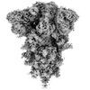 36724MC 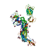 8jymC 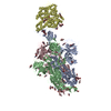 8jynC 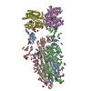 8jyoC 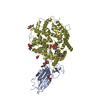 8jypC M: map data used to model this data C: citing same article ( |
|---|---|
| Similar structure data |
-Links
 PDBj
PDBj


- Assembly
| Deposited unit | 
|
|---|---|
| 1 |
|
| 2 |
|
| 3 |
|
| Symmetry | Point symmetry: (Schoenflies symbol: C3 (3 fold cyclic)) |
-Components
| #1: Protein | Spike protein / S glycoprotein / E2 / Peplomer protein Mass: 138086.094 Da / Num. of mol.: 1 Mutation: F817P, A892P, A899P, A942P, K986P, V987P, R682G, R683S, R685G Source method: isolated from a genetically manipulated source Source: (gene. exp.) Severe acute respiratory syndrome coronavirus 2Gene: S, 2 / Production host:  Homo sapiens (human) / References: UniProt: P0DTC2 Homo sapiens (human) / References: UniProt: P0DTC2 | ||||
|---|---|---|---|---|---|
| #2: Polysaccharide | / Mass: 424.401 Da / Num. of mol.: 3 Source method: isolated from a genetically manipulated source #3: Sugar | ChemComp-NAG / N-Acetylglucosamine  Type: D-saccharide, beta linking / Mass: 221.208 Da / Num. of mol.: 12 Type: D-saccharide, beta linking / Mass: 221.208 Da / Num. of mol.: 12Source method: isolated from a genetically manipulated source Formula: C8H15NO6 / Feature type: SUBJECT OF INVESTIGATION Has ligand of interest | Y | |
-Experimental details
-Experiment
| Experiment | Method: ELECTRON MICROSCOPY |
|---|---|
| EM experiment | Aggregation state: PARTICLE / 3D reconstruction method: single particle reconstruction |
- Sample preparation
| Component | Name: SARS-CoV-2 XBB1.5 spike glycoprotein / Type: COMPLEX / Entity ID: #1 / Source: RECOMBINANT | ||||||||||||||||||||
|---|---|---|---|---|---|---|---|---|---|---|---|---|---|---|---|---|---|---|---|---|---|
| Molecular weight | Value: 0.42 MDa / Experimental value: NO | ||||||||||||||||||||
| Source (natural) | Organism: Severe acute respiratory syndrome coronavirus 2 | ||||||||||||||||||||
| Source (recombinant) | Organism: Homo sapiens (human) | ||||||||||||||||||||
| Details of virus | Empty: YES / Enveloped: YES | ||||||||||||||||||||
| Natural host | Organism: Homo sapiens | ||||||||||||||||||||
| Buffer solution | pH: 7.4 | ||||||||||||||||||||
| Buffer component |
| ||||||||||||||||||||
| Specimen | Conc.: 1.2 mg/ml / Embedding applied: NO / Shadowing applied: NO / Staining applied: NO / Vitrification applied: YES | ||||||||||||||||||||
| Specimen support | Grid material: COPPER / Grid mesh size: 300 divisions/in. / Grid type: Quantifoil R2/2 | ||||||||||||||||||||
| Vitrification | Instrument: FEI VITROBOT MARK IV / Cryogen name: ETHANE / Humidity: 100 % / Chamber temperature: 291 K / Details: blotting time 5 s and blotting force 5. |
- Electron microscopy imaging
| Experimental equipment |  Model: Titan Krios / Image courtesy: FEI Company |
|---|---|
| Microscopy | Model: TFS KRIOS |
| Electron gun | Electron source: FIELD EMISSION GUN / Accelerating voltage: 300 kV / Illumination mode: FLOOD BEAM |
| Electron lens | Mode: BRIGHT FIELDBright-field microscopy / Nominal magnification: 130000 X / Nominal defocus max: 1800 nm / Nominal defocus min: 800 nm |
| Specimen holder | Cryogen: NITROGEN / Specimen holder model: FEI TITAN KRIOS AUTOGRID HOLDER |
| Image recording | Average exposure time: 1.5 sec. / Electron dose: 50.4 e/Å2 / Film or detector model: GATAN K3 (6k x 4k) / Num. of real images: 3522 |
| EM imaging optics | Energyfilter name: GIF Bioquantum / Energyfilter slit width: 20 eV |
| Image scans | Width: 5760 / Height: 4092 |
- Processing
| EM software |
| ||||||||||||||||||||||||||||||||||||||||
|---|---|---|---|---|---|---|---|---|---|---|---|---|---|---|---|---|---|---|---|---|---|---|---|---|---|---|---|---|---|---|---|---|---|---|---|---|---|---|---|---|---|
| CTF correction | Type: PHASE FLIPPING AND AMPLITUDE CORRECTION | ||||||||||||||||||||||||||||||||||||||||
| Particle selection | Num. of particles selected: 1201208 | ||||||||||||||||||||||||||||||||||||||||
| Symmetry | Point symmetry: C3 (3 fold cyclic) | ||||||||||||||||||||||||||||||||||||||||
| 3D reconstruction | Resolution: 2.59 Å / Resolution method: FSC 0.143 CUT-OFF / Num. of particles: 84161 / Algorithm: FOURIER SPACE / Num. of class averages: 1 / Symmetry type: POINT | ||||||||||||||||||||||||||||||||||||||||
| Atomic model building | Protocol: RIGID BODY FIT / Space: REAL | ||||||||||||||||||||||||||||||||||||||||
| Atomic model building | PDB-ID: 8IOS Accession code: 8IOS / Source name: PDB / Type: experimental model | ||||||||||||||||||||||||||||||||||||||||
| Refine LS restraints |
|

