 Movie
Movie Controller


+ Open data

- Basic information
| Entry | Database: PDB / ID: 8jqe | ||||||
|---|---|---|---|---|---|---|---|
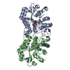
| Title | Structure of CmCBDA in complex with Mn2+ and glycerol | ||||||
 Components Components | YdjC family protein | ||||||
 Keywords Keywords |  HYDROLASE / CmCBDA / N-acetylglucosamine deacetylase / mentalloenzyme / enzyme engineering HYDROLASE / CmCBDA / N-acetylglucosamine deacetylase / mentalloenzyme / enzyme engineering | ||||||
| Function / homology | Carbohydrate deacetylase YdjC-like / YdjC-like protein / Glycoside hydrolase/deacetylase, beta/alpha-barrel / carbohydrate metabolic process / metal ion binding / : / PHOSPHATE ION / YdjC family protein Function and homology information Function and homology information | ||||||
| Biological species |  Cyclobacterium marinum (bacteria) Cyclobacterium marinum (bacteria) | ||||||
| Method | X-RAY DIFFRACTION / SYNCHROTRON / MOLECULAR REPLACEMENT / Resolution: 2.31 Å | ||||||
 Authors Authors | Li, X. | ||||||
| Funding support | 1items
| ||||||
 Citation Citation | Journal: J.Agric.Food Chem. / Year: 2024 Title: Structural Insights into the Catalytic Activity of Cyclobacterium marinum N -Acetylglucosamine Deacetylase. Authors: Hu, S. / Xu, L. / Xie, C. / Hong, J. | ||||||
| History |
|
- Structure visualization
| Structure viewer | Molecule: MolmilJmol/JSmol |
|---|
- Downloads & links
-Download
| PDBx/mmCIF format | 8jqe.cif.gz | 251.1 KB | Display | PDBx/mmCIF format |
|---|---|---|---|---|
| PDB format | pdb8jqe.ent.gz | 202.2 KB | Display | PDB format |
| PDBx/mmJSON format | 8jqe.json.gz | Tree view | PDBx/mmJSON format | |
| Others |  Other downloads Other downloads |
-Validation report
| Arichive directory | https://data.pdbj.org/pub/pdb/validation_reports/jq/8jqeftp://data.pdbj.org/pub/pdb/validation_reports/jq/8jqe | HTTPS FTP |
|---|
-Related structure data
-Links
 PDBj
PDBj
- Assembly
| Deposited unit | 
| ||||||||
|---|---|---|---|---|---|---|---|---|---|
| 1 |
| ||||||||
| Unit cell |
|
-Components
| #1: Protein | Mass: 35334.184 Da / Num. of mol.: 2 Source method: isolated from a genetically manipulated source Source: (gene. exp.) Cyclobacterium marinum (bacteria) / Gene: Cycma_4778 / Production host: Escherichia coli (E. coli) / References: UniProt: G0J5L4#2: Chemical | Glycerol  Mass: 92.094 Da / Num. of mol.: 2 / Source method: obtained synthetically / Formula: C3H8O3 / Feature type: SUBJECT OF INVESTIGATION Mass: 92.094 Da / Num. of mol.: 2 / Source method: obtained synthetically / Formula: C3H8O3 / Feature type: SUBJECT OF INVESTIGATION#3: Chemical |   Mass: 54.938 Da / Num. of mol.: 2 / Source method: obtained synthetically / Formula: Mn / Feature type: SUBJECT OF INVESTIGATION Mass: 54.938 Da / Num. of mol.: 2 / Source method: obtained synthetically / Formula: Mn / Feature type: SUBJECT OF INVESTIGATION#4: Chemical | Phosphate  Mass: 94.971 Da / Num. of mol.: 2 / Source method: obtained synthetically / Formula: PO4 / Feature type: SUBJECT OF INVESTIGATION Mass: 94.971 Da / Num. of mol.: 2 / Source method: obtained synthetically / Formula: PO4 / Feature type: SUBJECT OF INVESTIGATION#5: Water | ChemComp-HOH / | Water Mass: 18.015 Da / Num. of mol.: 124 / Source method: isolated from a natural source / Formula: H2O Mass: 18.015 Da / Num. of mol.: 124 / Source method: isolated from a natural source / Formula: H2OHas ligand of interest | Y | |
|---|
-Experimental details
-Experiment
| Experiment | Method: X-RAY DIFFRACTION / Number of used crystals: 1 |
|---|
- Sample preparation
| Crystal | Density Matthews: 2.35 Å3/Da / Density % sol: 47.71 % |
|---|---|
| Crystal grow | Temperature: 293 K / Method: vapor diffusion, sitting drop Details: 0.04M potassium dihydrogen phosphate, 16% PEG 8000, 20% Glycerol, 10mM MnCl2, 10 mM GlcNAc |
-Data collection
| Diffraction | Mean temperature: 100 K / Serial crystal experiment: N |
|---|---|
| Diffraction source | Source: SYNCHROTRON / Site: SSRF  / Beamline: BL19U1 / Wavelength: 0.9778 Å / Beamline: BL19U1 / Wavelength: 0.9778 Å |
| Detector | Type: DECTRIS PILATUS3 6M / Detector: PIXEL / Date: Sep 20, 2018 |
| Radiation | Protocol: SINGLE WAVELENGTH / Monochromatic (M) / Laue (L): M / Scattering type: x-ray |
| Radiation wavelength | Wavelength: 0.9778 Å / Relative weight: 1 |
| Reflection | Resolution: 2.3→40 Å / Num. obs: 28934 / % possible obs: 99.7 % / Redundancy: 4.2 % / Biso Wilson estimate: 32.62 Å2 / Rmerge(I) obs: 0.125 / Net I/σ(I): 13.49 |
| Reflection shell | Resolution: 2.3→2.34 Å / Rmerge(I) obs: 0.522 / Num. unique obs: 1414 |
- Processing
| Software |
| |||||||||||||||||||||||||||||||||||||||||||||||||||||||||||||||||||||||||||||
|---|---|---|---|---|---|---|---|---|---|---|---|---|---|---|---|---|---|---|---|---|---|---|---|---|---|---|---|---|---|---|---|---|---|---|---|---|---|---|---|---|---|---|---|---|---|---|---|---|---|---|---|---|---|---|---|---|---|---|---|---|---|---|---|---|---|---|---|---|---|---|---|---|---|---|---|---|---|---|
| Refinement | Method to determine structure: MOLECULAR REPLACEMENT / Resolution: 2.31→34.99 Å / SU ML: 0.2 / Cross valid method: THROUGHOUT / σ(F): 1.38 / Phase error: 20.56 / Stereochemistry target values: ML
| |||||||||||||||||||||||||||||||||||||||||||||||||||||||||||||||||||||||||||||
| Solvent computation | Shrinkage radii: 0.6 Å / VDW probe radii: 1 Å / Solvent model: FLAT BULK SOLVENT MODEL | |||||||||||||||||||||||||||||||||||||||||||||||||||||||||||||||||||||||||||||
| Refinement step | Cycle: LAST / Resolution: 2.31→34.99 Å
| |||||||||||||||||||||||||||||||||||||||||||||||||||||||||||||||||||||||||||||
| Refine LS restraints |
| |||||||||||||||||||||||||||||||||||||||||||||||||||||||||||||||||||||||||||||
| LS refinement shell |
| |||||||||||||||||||||||||||||||||||||||||||||||||||||||||||||||||||||||||||||
| Refinement TLS params. | Method: refined / Refine-ID: X-RAY DIFFRACTION
| |||||||||||||||||||||||||||||||||||||||||||||||||||||||||||||||||||||||||||||
| Refinement TLS group |
|
