 Movie
Movie Controller

[English] 日本語
 Yorodumi
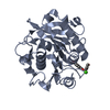
- PDB-8jmp: Structure of a leaf-branch compost cutinase, ICCG in complex with... -
+ Open data

- Basic information
| Entry | Database: PDB / ID: 8jmp | ||||||||||||
|---|---|---|---|---|---|---|---|---|---|---|---|---|---|
| Title | Structure of a leaf-branch compost cutinase, ICCG in complex with 1,4-butanediol terephthalate | ||||||||||||
 Components Components | Leaf-branch compost cutinase | ||||||||||||
 Keywords Keywords |  HYDROLASE / PETase / cutinase / enzyme engineering / PBAT degradation HYDROLASE / PETase / cutinase / enzyme engineering / PBAT degradation | ||||||||||||
| Function / homology |  Function and homology information Function and homology informationpoly(ethylene terephthalate) hydrolase / acetylesterase activity / cutinase activity / cutinase / extracellular regionSimilarity search - Function | ||||||||||||
| Biological species |  unidentified prokaryotic organism (environmental samples) unidentified prokaryotic organism (environmental samples) | ||||||||||||
| Method | X-RAY DIFFRACTION / MOLECULAR REPLACEMENT / Resolution: 1.9 Å | ||||||||||||
 Authors Authors | Yang, Y. / Xue, T. / Zheng, Y. / Cheng, S. / Guo, R.-T. / Chen, C.-C. | ||||||||||||
| Funding support |  China, 3items China, 3items
| ||||||||||||
 Citation Citation | Journal: J Hazard Mater / Year: 2023 Title: Remodeling the polymer-binding cavity to improve the efficacy of PBAT-degrading enzyme. Authors: Yang, Y. / Cheng, S. / Zheng, Y. / Xue, T. / Huang, J.W. / Zhang, L. / Yang, Y. / Guo, R.T. / Chen, C.C. | ||||||||||||
| History |
|
- Structure visualization
| Structure viewer | Molecule: MolmilJmol/JSmol |
|---|
- Downloads & links
-Download
| PDBx/mmCIF format | 8jmp.cif.gz | 129.5 KB | Display | PDBx/mmCIF format |
|---|---|---|---|---|
| PDB format | pdb8jmp.ent.gz | 96.3 KB | Display | PDB format |
| PDBx/mmJSON format | 8jmp.json.gz | Tree view | PDBx/mmJSON format | |
| Others |  Other downloads Other downloads |
-Validation report
| Arichive directory | https://data.pdbj.org/pub/pdb/validation_reports/jm/8jmpftp://data.pdbj.org/pub/pdb/validation_reports/jm/8jmp | HTTPS FTP |
|---|
-Related structure data
-Links
 PDBj
PDBj
- Assembly
| Deposited unit | 
| ||||||||
|---|---|---|---|---|---|---|---|---|---|
| 1 | 
| ||||||||
| 2 | 
| ||||||||
| Unit cell |
|
-Components
| #1: Protein | Mass: 27836.219 Da / Num. of mol.: 2 Source method: isolated from a genetically manipulated source Source: (gene. exp.) unidentified prokaryotic organism (environmental samples)Production host: Escherichia coli BL21(DE3) (bacteria)References: UniProt: G9BY57, cutinase, poly(ethylene terephthalate) hydrolase#2: Chemical | ChemComp-CA / |   Mass: 40.078 Da / Num. of mol.: 1 / Source method: obtained synthetically / Formula: Ca Mass: 40.078 Da / Num. of mol.: 1 / Source method: obtained synthetically / Formula: Ca#3: Chemical | ChemComp-EMX / |   Mass: 386.352 Da / Num. of mol.: 1 / Source method: obtained synthetically / Formula: C20H18O8 / Feature type: SUBJECT OF INVESTIGATION Mass: 386.352 Da / Num. of mol.: 1 / Source method: obtained synthetically / Formula: C20H18O8 / Feature type: SUBJECT OF INVESTIGATION#4: Water | ChemComp-HOH / | Water Mass: 18.015 Da / Num. of mol.: 667 / Source method: isolated from a natural source / Formula: H2O Mass: 18.015 Da / Num. of mol.: 667 / Source method: isolated from a natural source / Formula: H2OHas ligand of interest | Y | |
|---|
-Experimental details
-Experiment
| Experiment | Method: X-RAY DIFFRACTION / Number of used crystals: 1 |
|---|
- Sample preparation
| Crystal | Density Matthews: 2.42 Å3/Da / Density % sol: 49.19 % |
|---|---|
| Crystal grow | Temperature: 298 K / Method: evaporation Details: 20% v/v PEG 8000 and 0.1 M sodium cacodylate, pH 6.5 |
-Data collection
| Diffraction | Mean temperature: 100 K / Serial crystal experiment: N |
|---|---|
| Diffraction source | Source: LIQUID ANODE / Type: BRUKER METALJET / Wavelength: 1.34138 Å |
| Detector | Type: Bruker PHOTON II / Detector: PIXEL / Date: Apr 28, 2023 |
| Radiation | Protocol: SINGLE WAVELENGTH / Monochromatic (M) / Laue (L): M / Scattering type: x-ray |
| Radiation wavelength | Wavelength: 1.34138 Å / Relative weight: 1 |
| Reflection | Resolution: 1.9→36.94 Å / Num. obs: 78612 / % possible obs: 99.4 % / Redundancy: 9.23 % / CC1/2: 1 / Rmerge(I) obs: 0.058 / Net I/σ(I): 25.33 |
| Reflection shell | Resolution: 1.9→1.93 Å / Rmerge(I) obs: 0.1439 / Mean I/σ(I) obs: 6.26 / Num. unique obs: 1906 / CC1/2: 0.989 |
- Processing
| Software |
| ||||||||||||||||||||||||||||||||||||||||||||||||||||||||||||||||||||||||||||||||||||||||||||||||||||||||||||||||||||||||||||||||||||||||||||||||||||||||||||||||||||||||||||||||||||||||||||||||||||
|---|---|---|---|---|---|---|---|---|---|---|---|---|---|---|---|---|---|---|---|---|---|---|---|---|---|---|---|---|---|---|---|---|---|---|---|---|---|---|---|---|---|---|---|---|---|---|---|---|---|---|---|---|---|---|---|---|---|---|---|---|---|---|---|---|---|---|---|---|---|---|---|---|---|---|---|---|---|---|---|---|---|---|---|---|---|---|---|---|---|---|---|---|---|---|---|---|---|---|---|---|---|---|---|---|---|---|---|---|---|---|---|---|---|---|---|---|---|---|---|---|---|---|---|---|---|---|---|---|---|---|---|---|---|---|---|---|---|---|---|---|---|---|---|---|---|---|---|---|---|---|---|---|---|---|---|---|---|---|---|---|---|---|---|---|---|---|---|---|---|---|---|---|---|---|---|---|---|---|---|---|---|---|---|---|---|---|---|---|---|---|---|---|---|---|---|---|---|
| Refinement | Method to determine structure: MOLECULAR REPLACEMENT / Resolution: 1.9→31.62 Å / SU ML: 0.15 / Cross valid method: NONE / σ(F): 1.35 / Phase error: 19.6 / Stereochemistry target values: ML
| ||||||||||||||||||||||||||||||||||||||||||||||||||||||||||||||||||||||||||||||||||||||||||||||||||||||||||||||||||||||||||||||||||||||||||||||||||||||||||||||||||||||||||||||||||||||||||||||||||||
| Solvent computation | Shrinkage radii: 0.9 Å / VDW probe radii: 1.11 Å / Solvent model: FLAT BULK SOLVENT MODEL | ||||||||||||||||||||||||||||||||||||||||||||||||||||||||||||||||||||||||||||||||||||||||||||||||||||||||||||||||||||||||||||||||||||||||||||||||||||||||||||||||||||||||||||||||||||||||||||||||||||
| Refinement step | Cycle: LAST / Resolution: 1.9→31.62 Å
| ||||||||||||||||||||||||||||||||||||||||||||||||||||||||||||||||||||||||||||||||||||||||||||||||||||||||||||||||||||||||||||||||||||||||||||||||||||||||||||||||||||||||||||||||||||||||||||||||||||
| Refine LS restraints |
| ||||||||||||||||||||||||||||||||||||||||||||||||||||||||||||||||||||||||||||||||||||||||||||||||||||||||||||||||||||||||||||||||||||||||||||||||||||||||||||||||||||||||||||||||||||||||||||||||||||
| LS refinement shell |
|
