[English] 日本語
Yorodumi
- PDB-8jkz: Cryo-EM structure of the prokaryotic SPARSA system complex -

+
Open data


ID or keywords:

Loading...

-
Basic information

Entry
Database: PDB / ID: 8jkz
TitleCryo-EM structure of the prokaryotic SPARSA system complex
Components
  • Piwi domain proteinPiwi
  • Sir2 superfamily protein
KeywordsANTIVIRAL PROTEIN / SPARSA antiviral system / NADase / Argonaute proteins
Function / homologySIR2-like domain / SIR2-like domain / DHS-like NAD/FAD-binding domain superfamily / Ribonuclease H superfamily / nucleic acid binding / Ribonuclease H-like superfamily / NICOTINAMIDE-ADENINE-DINUCLEOTIDE / Piwi domain protein / Sir2 superfamily protein
Function and homology information
Biological speciesGeobacter sulfurreducens (bacteria)
MethodELECTRON MICROSCOPY / single particle reconstruction / cryo EM / Resolution: 3.6 Å
AuthorsXu, X. / Zhen, X. / Long, F.
Funding support China, 3items
OrganizationGrant numberCountry
Ministry of Science and Technology (MoST, China)2021YFA0909500 China
Ministry of Science and Technology (MoST, China)2018YFA0900400 China
Ministry of Education (MoE, China)2042019kf0185 China
Citation
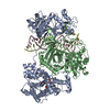
Journal: Nat Commun / Year: 2024
Title: Structural basis of antiphage immunity generated by a prokaryotic Argonaute-associated SPARSA system.
Authors: Xiangkai Zhen / Xiaolong Xu / Le Ye / Song Xie / Zhijie Huang / Sheng Yang / Yanhui Wang / Jinyu Li / Feng Long / Songying Ouyang /
Abstract: Argonaute (Ago) proteins are ubiquitous across all kingdoms of life. Eukaryotic Agos (eAgos) use small RNAs to recognize transcripts for RNA silencing in eukaryotes. In contrast, the functions of ...Argonaute (Ago) proteins are ubiquitous across all kingdoms of life. Eukaryotic Agos (eAgos) use small RNAs to recognize transcripts for RNA silencing in eukaryotes. In contrast, the functions of prokaryotic counterparts (pAgo) are less well known. Recently, short pAgos in conjunction with the associated TIR or Sir2 (SPARTA or SPARSA) were found to serve as antiviral systems to combat phage infections. Herein, we present the cryo-EM structures of nicotinamide adenine dinucleotide (NAD)-bound SPARSA with and without nucleic acids at resolutions of 3.1 Å and 3.6 Å, respectively. Our results reveal that the APAZ (Analogue of PAZ) domain and the short pAgo form a featured architecture similar to the long pAgo to accommodate nucleic acids. We further identified the key residues for NAD binding and elucidated the structural basis for guide RNA and target DNA recognition. Using structural comparisons, molecular dynamics simulations, and biochemical experiments, we proposed a putative mechanism for NAD hydrolysis in which an H186 loop mediates nucleophilic attack by catalytic water molecules. Overall, our study provides mechanistic insight into the antiphage role of the SPARSA system.
History
DepositionJun 2, 2023Deposition site: PDBJ / Processing site: PDBJ
Revision 1.0Jan 24, 2024Provider: repository / Type: Initial release

-
Structure visualization

Structure viewerMolecule:
MolmilJmol/JSmol

Downloads & links

-
Assembly

Deposited unit
A: Sir2 superfamily protein
B: Piwi domain protein
hetero molecules


Theoretical massNumber of molelcules
Total (without water)120,2573
Polymers119,5942
Non-polymers6631
Water0
1


  • Idetical with deposited unit
  • defined by author
  • Evidence: electron microscopy, not applicable
TypeNameSymmetry operationNumber
identity operation1_5551

-
Components

#1: Protein Sir2 superfamily protein


Mass: 66268.500 Da / Num. of mol.: 1
Source method: isolated from a genetically manipulated source
Source: (gene. exp.) Geobacter sulfurreducens (bacteria) / Gene: GSU1360 / Plasmid: pET21a / Production host: Escherichia coli BL21(DE3) (bacteria) / References: UniProt: Q74DF6
#2: Protein Piwi domain protein / Piwi


Mass: 53325.566 Da / Num. of mol.: 1
Source method: isolated from a genetically manipulated source
Source: (gene. exp.) Geobacter sulfurreducens (bacteria) / Gene: GSU1361 / Plasmid: pET21a / Production host: Escherichia coli BL21(DE3) (bacteria) / References: UniProt: Q74DF5
#3: Chemical ChemComp-NAD / NICOTINAMIDE-ADENINE-DINUCLEOTIDE / Nicotinamide adenine dinucleotide


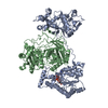
Mass: 663.425 Da / Num. of mol.: 1 / Source method: obtained synthetically / Formula: C21H27N7O14P2 / Feature type: SUBJECT OF INVESTIGATION / Comment: NAD*YM
Has ligand of interestY

-
Experimental details

-
Experiment

ExperimentMethod: ELECTRON MICROSCOPY
EM experimentAggregation state: PARTICLE / 3D reconstruction method: single particle reconstruction

-
Sample preparation

Component
IDNameTypeEntity IDParent-IDSource
1Sir2/AgoCOMPLEX#20RECOMBINANT
2Sir2Sirtuin 1COMPLEX#11RECOMBINANT
3AgoCOMPLEX#21RECOMBINANT
Molecular weight
IDEntity assembly-IDExperimental value
11NO
21NO
31NO
Source (natural)
IDEntity assembly-IDOrganismNcbi tax-ID
21Geobacter sulfurreducens PCA (bacteria)243231
32Geobacter sulfurreducens PCA (bacteria)243231
Source (recombinant)
IDEntity assembly-IDOrganismNcbi tax-IDStrain
21Escherichia coli (E. coli)562BL21 (DE3)
32Escherichia coli (E. coli)562BL21 (DE3)
Buffer solutionpH: 8
Buffer component
IDConc.NameFormulaBuffer-ID
150 mMTris bufferTris-HClTris1
2150 mMsodium chlorideNaClSodium chloride1
SpecimenConc.: 2 mg/ml / Embedding applied: NO / Shadowing applied: NO / Staining applied: NO / Vitrification applied: YES
Specimen supportGrid material: COPPER / Grid mesh size: 300 divisions/in. / Grid type: Quantifoil R1.2/1.3
VitrificationInstrument: FEI VITROBOT MARK IV / Cryogen name: ETHANE / Humidity: 100 % / Chamber temperature: 281 K

-
Electron microscopy imaging

Experimental equipment
Model: Titan Krios / Image courtesy: FEI Company
MicroscopyModel: FEI TITAN KRIOS
Electron gunElectron source: FIELD EMISSION GUN / Accelerating voltage: 300 kV / Illumination mode: FLOOD BEAM
Electron lensMode: BRIGHT FIELDBright-field microscopy / Nominal defocus max: 2000 nm / Nominal defocus min: 1000 nm / Cs: 2.7 mm
Image recordingElectron dose: 50 e/Å2 / Film or detector model: GATAN K3 (6k x 4k)

-
Processing

EM software
IDNameVersionCategory
2EPUFEIimage acquisition
13PHENIX1.20.1_4487:model refinement
CTF correctionType: NONE
SymmetryPoint symmetry: C1 (asymmetric)
3D reconstructionResolution: 3.6 Å / Resolution method: FSC 0.143 CUT-OFF / Num. of particles: 46372 / Symmetry type: POINT
Atomic model buildingSource name: AlphaFold / Type: in silico model
Refine LS restraints
Refine-IDTypeDev idealNumber
ELECTRON MICROSCOPYf_bond_d0.0038654
ELECTRON MICROSCOPYf_angle_d0.59311744
ELECTRON MICROSCOPYf_dihedral_angle_d5.1231157
ELECTRON MICROSCOPYf_chiral_restr0.0411280
ELECTRON MICROSCOPYf_plane_restr0.0031505

+
About Yorodumi

-
News

-
Feb 9, 2022. New format data for meta-information of EMDB entries

New format data for meta-information of EMDB entries

  • Version 3 of the EMDB header file is now the official format.
  • The previous official version 1.9 will be removed from the archive.

Related info.:EMDB header

External links:wwPDB to switch to version 3 of the EMDB data model

-
Aug 12, 2020. Covid-19 info

Covid-19 info

URL: https://pdbj.org/emnavi/covid19.php

New page: Covid-19 featured information page in EM Navigator.

Related info.:Covid-19 info / Mar 5, 2020. Novel coronavirus structure data

+
Mar 5, 2020. Novel coronavirus structure data

Novel coronavirus structure data

Related info.:Yorodumi Speices / Aug 12, 2020. Covid-19 info

External links:COVID-19 featured content - PDBj / Molecule of the Month (242):Coronavirus Proteases

+
Jan 31, 2019. EMDB accession codes are about to change! (news from PDBe EMDB page)

EMDB accession codes are about to change! (news from PDBe EMDB page)

  • The allocation of 4 digits for EMDB accession codes will soon come to an end. Whilst these codes will remain in use, new EMDB accession codes will include an additional digit and will expand incrementally as the available range of codes is exhausted. The current 4-digit format prefixed with “EMD-” (i.e. EMD-XXXX) will advance to a 5-digit format (i.e. EMD-XXXXX), and so on. It is currently estimated that the 4-digit codes will be depleted around Spring 2019, at which point the 5-digit format will come into force.
  • The EM Navigator/Yorodumi systems omit the EMD- prefix.

Related info.:Q: What is EMD? / ID/Accession-code notation in Yorodumi/EM Navigator

External links:EMDB Accession Codes are Changing Soon! / Contact to PDBj

+
Jul 12, 2017. Major update of PDB

Major update of PDB

  • wwPDB released updated PDB data conforming to the new PDBx/mmCIF dictionary.
  • This is a major update changing the version number from 4 to 5, and with Remediation, in which all the entries are updated.
  • In this update, many items about electron microscopy experimental information are reorganized (e.g. em_software).
  • Now, EM Navigator and Yorodumi are based on the updated data.

External links:wwPDB Remediation / Enriched Model Files Conforming to OneDep Data Standards Now Available in the PDB FTP Archive

-
Yorodumi

Thousand views of thousand structures

  • Yorodumi is a browser for structure data from EMDB, PDB, SASBDB, etc.
  • This page is also the successor to EM Navigator detail page, and also detail information page/front-end page for Omokage search.
  • The word "yorodu" (or yorozu) is an old Japanese word meaning "ten thousand". "mi" (miru) is to see.

Related info.:EMDB / PDB / SASBDB / Comparison of 3 databanks / Yorodumi Search / Aug 31, 2016. New EM Navigator & Yorodumi / Yorodumi Papers / Jmol/JSmol / Function and homology information / Changes in new EM Navigator and Yorodumi

Read more