 Movie
Movie Controller


+ Open data

- Basic information
| Entry | Database: PDB / ID: 8jes | ||||||
|---|---|---|---|---|---|---|---|
| Title | Cryo-EM structure of DltB homo-tetramer | ||||||
 Components Components | Teichoic acid D-alanyltransferase | ||||||
 Keywords Keywords |  MEMBRANE PROTEIN / channel / anti-virulence / MBOAT / DltB MEMBRANE PROTEIN / channel / anti-virulence / MBOAT / DltB | ||||||
| Function / homology |  Function and homology information Function and homology informationlipoteichoic acid biosynthetic process / acyltransferase activity / Transferases; Acyltransferases; Transferring groups other than aminoacyl groups / plasma membraneSimilarity search - Function | ||||||
| Biological species |  Streptococcus thermophilus LMG 18311 (bacteria) Streptococcus thermophilus LMG 18311 (bacteria) | ||||||
| Method | ELECTRON MICROSCOPY / single particle reconstruction / cryo EM / Resolution: 3.42 Å | ||||||
 Authors Authors | Zhang, P. / Liu, Z. | ||||||
| Funding support | 1items
| ||||||
 Citation Citation | Journal: Nat Commun / Year: 2024 Title: Structural insights into the transporting and catalyzing mechanism of DltB in LTA D-alanylation. Authors: Pingfeng Zhang / Zheng Liu /  Abstract: DltB, a model member of the Membrane-Bound O-AcylTransferase (MBOAT) superfamily, plays a crucial role in D-alanylation of the lipoteichoic acid (LTA), a significant component of the cell wall of ...DltB, a model member of the Membrane-Bound O-AcylTransferase (MBOAT) superfamily, plays a crucial role in D-alanylation of the lipoteichoic acid (LTA), a significant component of the cell wall of gram-positive bacteria. This process stabilizes the cell wall structure, influences bacterial virulence, and modulates the host immune response. Despite its significance, the role of DltB is not well understood. Through biochemical analysis and cryo-EM imaging, we discover that Streptococcus thermophilus DltB forms a homo-tetramer on the cell membrane. We further visualize DltB in an apo form, in complex with DltC, and in complex with its inhibitor amsacrine (m-AMSA). Each tetramer features a central hole. The C-tunnel of each protomer faces the intratetramer interface and provides access to the periphery membrane. Each protomer binds a DltC without changing the tetrameric organization. A phosphatidylglycerol (PG) molecule in the substrate-binding site may serve as an LTA carrier. The inhibitor m-AMSA bound to the L-tunnel of each protomer blocks the active site. The tetrameric organization of DltB provides a scaffold for catalyzing D-alanyl transfer and regulating the channel opening and closing. Our findings unveil DltB's dual function in the D-alanylation pathway, and provide insight for targeting DltB as a anti-virulence antibiotic. | ||||||
| History |
|
- Structure visualization
| Structure viewer | Molecule: MolmilJmol/JSmol |
|---|
- Downloads & links
-Download
| PDBx/mmCIF format | 8jes.cif.gz | 474.4 KB | Display | PDBx/mmCIF format |
|---|---|---|---|---|
| PDB format | pdb8jes.ent.gz | 321.3 KB | Display | PDB format |
| PDBx/mmJSON format | 8jes.json.gz | Tree view | PDBx/mmJSON format | |
| Others |  Other downloads Other downloads |
-Validation report
| Arichive directory | https://data.pdbj.org/pub/pdb/validation_reports/je/8jesftp://data.pdbj.org/pub/pdb/validation_reports/je/8jes | HTTPS FTP |
|---|
-Related structure data
| Related structure data |  36194MC 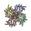 8jemC 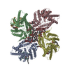 8jf2C C: citing same article ( M: map data used to model this data |
|---|---|
| Similar structure data |


-Links
 PDBj
PDBj
- Assembly
| Deposited unit | 
|
|---|---|
| 1 |
|
-Components
| #1: Protein | Mass: 51780.027 Da / Num. of mol.: 4 Source method: isolated from a genetically manipulated source Source: (gene. exp.) Streptococcus thermophilus LMG 18311 (bacteria)Strain: LMG 18311 / Gene: dltB, stu0762 / Production host: Escherichia coli BL21(DE3) (bacteria) / Variant (production host): C41References: UniProt: Q5M4V4, Transferases; Acyltransferases; Transferring groups other than aminoacyl groups#2: Chemical | ChemComp-PGT / ( Phosphatidylglycerol  Mass: 751.023 Da / Num. of mol.: 6 / Source method: obtained synthetically / Formula: C40H79O10P / Feature type: SUBJECT OF INVESTIGATION / Comment: phospholipid*YM Mass: 751.023 Da / Num. of mol.: 6 / Source method: obtained synthetically / Formula: C40H79O10P / Feature type: SUBJECT OF INVESTIGATION / Comment: phospholipid*YM#3: Sugar | ChemComp-LMT /   Type: D-saccharide / Mass: 510.615 Da / Num. of mol.: 35 / Source method: obtained synthetically / Formula: C24H46O11 / Comment: detergent*YM Type: D-saccharide / Mass: 510.615 Da / Num. of mol.: 35 / Source method: obtained synthetically / Formula: C24H46O11 / Comment: detergent*YM#4: Chemical | Diglyceride  Mass: 625.018 Da / Num. of mol.: 2 / Source method: obtained synthetically / Formula: C39H76O5 / Feature type: SUBJECT OF INVESTIGATION Mass: 625.018 Da / Num. of mol.: 2 / Source method: obtained synthetically / Formula: C39H76O5 / Feature type: SUBJECT OF INVESTIGATIONHas ligand of interest | Y | |
|---|
-Experimental details
-Experiment
| Experiment | Method: ELECTRON MICROSCOPY |
|---|---|
| EM experiment | Aggregation state: PARTICLE / 3D reconstruction method: single particle reconstruction |
- Sample preparation
| Component | Name: apo DltB tetramer in DDM. / Type: ORGANELLE OR CELLULAR COMPONENT / Entity ID: #1 / Source: RECOMBINANT |
|---|---|
| Molecular weight | Value: 48 kDa/nm / Experimental value: YES |
| Source (natural) | Organism: Streptococcus thermophilus (bacteria) / Strain: LMG 18311 |
| Source (recombinant) | Organism: Escherichia coli BL21(DE3) (bacteria) / Strain: C41(DE3) |
| Buffer solution | pH: 7.5 / Details: 20 mM Hopes-Na, pH7.5, 0.03% DDM |
| Buffer component | Conc.: 25 mM / Name: Hepes |
| Specimen | Conc.: 9 mg/ml / Embedding applied: NO / Shadowing applied: NO / Staining applied: NO / Vitrification applied: YES Details: The DltB protein was purified in DDM, the tetramer fractions from gel filtration column were concentrated to about 10 mg/ml. |
| Vitrification | Instrument: FEI VITROBOT MARK IV / Cryogen name: ETHANE / Humidity: 100 % / Chamber temperature: 277 K Details: After incubation on the grids at 277K under 100% humidity for 10 s, the grids were bloted for 3.0 s and then plunged frozen into liquid ethane cooled by liquid nitrogen using a Vitrobot. |
- Electron microscopy imaging
| Experimental equipment |  Model: Titan Krios / Image courtesy: FEI Company |
|---|---|
| Microscopy | Model: FEI TITAN KRIOS |
| Electron gun | Electron source: FIELD EMISSION GUN / Accelerating voltage: 300 kV / Illumination mode: FLOOD BEAM |
| Electron lens | Mode: BRIGHT FIELDBright-field microscopy / Nominal defocus max: 1800 nm / Nominal defocus min: 1100 nm |
| Image recording | Electron dose: 55 e/Å2 / Film or detector model: GATAN K3 BIOQUANTUM (6k x 4k) |
- Processing
| Software |
| ||||||||||||||||||||||||
|---|---|---|---|---|---|---|---|---|---|---|---|---|---|---|---|---|---|---|---|---|---|---|---|---|---|
| EM software | Name: SerialEM / Category: image acquisition | ||||||||||||||||||||||||
| CTF correction | Type: PHASE FLIPPING AND AMPLITUDE CORRECTION | ||||||||||||||||||||||||
| Particle selection | Num. of particles selected: 3538544 | ||||||||||||||||||||||||
| 3D reconstruction | Resolution: 3.42 Å / Resolution method: FSC 0.143 CUT-OFF / Num. of particles: 128038 / Symmetry type: POINT | ||||||||||||||||||||||||
| Atomic model building | Space: REAL | ||||||||||||||||||||||||
| Refinement | Cross valid method: NONE Stereochemistry target values: GeoStd + Monomer Library + CDL v1.2 | ||||||||||||||||||||||||
| Displacement parameters | Biso mean: 152.98 Å2 | ||||||||||||||||||||||||
| Refine LS restraints |
|
