 Movie
Movie Controller

[English] 日本語
 Yorodumi
- PDB-8jaa: Crystal structure of Mycobacterium tuberculosis LpqY in complex w... -
+ Open data

- Basic information
| Entry | Database: PDB / ID: 8jaa | |||||||||
|---|---|---|---|---|---|---|---|---|---|---|
| Title | Crystal structure of Mycobacterium tuberculosis LpqY in complex with trehalose analogue YB-04 | |||||||||
 Components Components | Trehalose-binding lipoprotein LpqY | |||||||||
 Keywords Keywords | SUGAR BINDING PROTEIN /  Trehalose / LpqY / Mycobacterium tuberculosis Trehalose / LpqY / Mycobacterium tuberculosis | |||||||||
| Function / homology |  Function and homology information Function and homology informationtrehalose transmembrane transporter activity / trehalose transport / biological process involved in interaction with host / ATP-binding cassette (ABC) transporter complex / periplasmic space / plasma membraneSimilarity search - Function | |||||||||
| Biological species |  Mycobacterium tuberculosis H37Rv (bacteria) Mycobacterium tuberculosis H37Rv (bacteria) | |||||||||
| Method | X-RAY DIFFRACTION / SYNCHROTRON / MOLECULAR REPLACEMENT / Resolution: 1.7 Å | |||||||||
 Authors Authors | Zhang, B. / Liang, J. / Rao, Z. | |||||||||
| Funding support |  China, 2items China, 2items
| |||||||||
 Citation Citation | Journal: Proc Natl Acad Sci U S A / Year: 2023 Title: Molecular recognition of trehalose and trehalose analogues by LpqY-SugABC. Authors: Jingxi Liang / Fengjiang Liu / Peng Xu / Wei Shangguan / Tianyu Hu / Shule Wang / Xiaolin Yang / Zhiqi Xiong / Xiuna Yang / Luke W Guddat / Biao Yu / Zihe Rao / Bing Zhang /  Abstract: Trehalose plays a crucial role in the survival and virulence of the deadly human pathogen (). The type I ATP-binding cassette (ABC) transporter LpqY-SugABC is the sole pathway for trehalose to enter ...Trehalose plays a crucial role in the survival and virulence of the deadly human pathogen (). The type I ATP-binding cassette (ABC) transporter LpqY-SugABC is the sole pathway for trehalose to enter . The substrate-binding protein, LpqY, which forms a stable complex with the translocator SugABC, recognizes and captures trehalose and its analogues in the periplasmic space, but the precise molecular mechanism for this process is still not well understood. This study reports a 3.02-Å cryoelectron microscopy structure of trehalose-bound LpqY-SugABC in the pretranslocation state, a crystal structure of LpqY in a closed form with trehalose bound and five crystal structures of LpqY in complex with different trehalose analogues. These structures, accompanied by substrate-stimulated ATPase activity data, reveal how LpqY recognizes and binds trehalose and its analogues, and highlight the flexibility in the substrate binding pocket of LpqY. These data provide critical insights into the design of trehalose analogues that could serve as potential molecular probe tools or as anti-TB drugs. | |||||||||
| History |
|
- Structure visualization
| Structure viewer | Molecule: MolmilJmol/JSmol |
|---|
- Downloads & links
-Download
| PDBx/mmCIF format | 8jaa.cif.gz | 105.3 KB | Display | PDBx/mmCIF format |
|---|---|---|---|---|
| PDB format | pdb8jaa.ent.gz | 75.9 KB | Display | PDB format |
| PDBx/mmJSON format | 8jaa.json.gz | Tree view | PDBx/mmJSON format | |
| Others |  Other downloads Other downloads |
-Validation report
| Arichive directory | https://data.pdbj.org/pub/pdb/validation_reports/ja/8jaaftp://data.pdbj.org/pub/pdb/validation_reports/ja/8jaa | HTTPS FTP |
|---|
-Related structure data
| Related structure data |  8ja7C 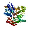 8ja8C  8ja9C 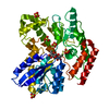 8jabC 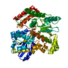 8jacC 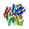 8jadC C: citing same article ( |
|---|---|
| Similar structure data |

-Links
 PDBj
PDBj


- Assembly
| Deposited unit | 
| ||||||||||||
|---|---|---|---|---|---|---|---|---|---|---|---|---|---|
| 1 |
| ||||||||||||
| Unit cell |
|
-Components
| #1: Protein | Mass: 49803.074 Da / Num. of mol.: 1 Source method: isolated from a genetically manipulated source Source: (gene. exp.) Mycobacterium tuberculosis H37Rv (bacteria)Gene: lpqY, Rv1235 Production host: Escherichia coli 'BL21-Gold(DE3)pLysS AG' (bacteria)References: UniProt: P9WGU9 | ||||||
|---|---|---|---|---|---|---|---|
| #2: Chemical | ChemComp-SO4 / Sulfate  Mass: 96.063 Da / Num. of mol.: 5 / Source method: obtained synthetically / Formula: SO4 Mass: 96.063 Da / Num. of mol.: 5 / Source method: obtained synthetically / Formula: SO4#3: Sugar |   Type: D-saccharide, alpha linking / Mass: 209.200 Da / Num. of mol.: 2 / Source method: obtained synthetically / Formula: C6H15N3O5 / Feature type: SUBJECT OF INVESTIGATION Type: D-saccharide, alpha linking / Mass: 209.200 Da / Num. of mol.: 2 / Source method: obtained synthetically / Formula: C6H15N3O5 / Feature type: SUBJECT OF INVESTIGATION#4: Water | ChemComp-HOH / | Water Mass: 18.015 Da / Num. of mol.: 256 / Source method: isolated from a natural source / Formula: H2O Mass: 18.015 Da / Num. of mol.: 256 / Source method: isolated from a natural source / Formula: H2OHas ligand of interest | Y | |
-Experimental details
-Experiment
| Experiment | Method: X-RAY DIFFRACTION / Number of used crystals: 1 |
|---|
- Sample preparation
| Crystal | Density Matthews: 2.76 Å3/Da / Density % sol: 55.41 % |
|---|---|
| Crystal grow | Temperature: 293 K / Method: vapor diffusion, sitting drop / pH: 7 Details: 50 mM Magnesium sulfate hydrate, 50 mM HEPES sodium pH 7.0 and 1.6 M Lithium sulfate monohydrate |
-Data collection
| Diffraction | Mean temperature: 100 K / Serial crystal experiment: N |
|---|---|
| Diffraction source | Source: SYNCHROTRON / Site: SSRF / Beamline: BL17U1 / Wavelength: 0.979 Å |
| Detector | Type: DECTRIS EIGER X 16M / Detector: PIXEL / Date: Dec 14, 2019 |
| Radiation | Protocol: SINGLE WAVELENGTH / Monochromatic (M) / Laue (L): M / Scattering type: x-ray |
| Radiation wavelength | Wavelength: 0.979 Å / Relative weight: 1 |
| Reflection | Resolution: 1.7→50 Å / Num. obs: 61357 / % possible obs: 99.1 % / Redundancy: 3.3 % / CC1/2: 0.997 / Net I/σ(I): 11.18 |
| Reflection shell | Resolution: 1.7→1.74 Å / Num. unique obs: 6042 / CC1/2: 0.882 |
- Processing
| Software |
| ||||||||||||||||||||||||
|---|---|---|---|---|---|---|---|---|---|---|---|---|---|---|---|---|---|---|---|---|---|---|---|---|---|
| Refinement | Method to determine structure: MOLECULAR REPLACEMENT / Resolution: 1.7→26.57 Å / Cross valid method: THROUGHOUT Stereochemistry target values: GeoStd + Monomer Library + CDL v1.2
| ||||||||||||||||||||||||
| Displacement parameters | Biso mean: 33.21 Å2 | ||||||||||||||||||||||||
| Refinement step | Cycle: LAST / Resolution: 1.7→26.57 Å
| ||||||||||||||||||||||||
| Refine LS restraints |
|
