 Movie
Movie Controller

[English] 日本語
 Yorodumi
- PDB-8j7r: Cryo-EM structure of the J-K-St region of EMCV IRES in complex wi... -
+ Open data

- Basic information
| Entry | Database: PDB / ID: 8j7r | |||||||||
|---|---|---|---|---|---|---|---|---|---|---|
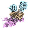
| Title | Cryo-EM structure of the J-K-St region of EMCV IRES in complex with eIF4G-HEAT1 and eIF4A (J-K-St/eIF4G focused) | |||||||||
 Components Components |
| |||||||||
 Keywords Keywords |  TRANSLATION / Translation initiation factors / RNA binding protein TRANSLATION / Translation initiation factors / RNA binding protein | |||||||||
| Function / homology |  Function and homology information Function and homology informationpositive regulation of eukaryotic translation initiation factor 4F complex assembly / positive regulation of mRNA cap binding / positive regulation of translation in response to endoplasmic reticulum stress / cap-dependent translational initiation / macromolecule biosynthetic process / Activation of the mRNA upon binding of the cap-binding complex and eIFs, and subsequent binding to 43S / eukaryotic initiation factor 4E binding / regulation of cellular response to stress / eukaryotic translation initiation factor 4F complex / Z-decay: degradation of maternal mRNAs by zygotically expressed factors ...positive regulation of eukaryotic translation initiation factor 4F complex assembly / positive regulation of mRNA cap binding / positive regulation of translation in response to endoplasmic reticulum stress / cap-dependent translational initiation / macromolecule biosynthetic process / Activation of the mRNA upon binding of the cap-binding complex and eIFs, and subsequent binding to 43S / eukaryotic initiation factor 4E binding / regulation of cellular response to stress / eukaryotic translation initiation factor 4F complex / Z-decay: degradation of maternal mRNAs by zygotically expressed factors / translation factor activity, RNA binding / Deadenylation of mRNA / miRNA-mediated gene silencing by inhibition of translation / M-decay: degradation of maternal mRNAs by maternally stored factors / positive regulation of protein localization to cell periphery / regulation of translational initiation / Ribosomal scanning and start codon recognition / Translation initiation complex formation / negative regulation of peptidyl-threonine phosphorylation / mTORC1-mediated signalling / cellular response to nutrient levels / regulation of presynapse assembly / Nonsense Mediated Decay (NMD) independent of the Exon Junction Complex (EJC) / GTP hydrolysis and joining of the 60S ribosomal subunit / L13a-mediated translational silencing of Ceruloplasmin expression / positive regulation of G1/S transition of mitotic cell cycle / behavioral fear response / Nonsense Mediated Decay (NMD) enhanced by the Exon Junction Complex (EJC) / translation initiation factor binding / energy homeostasis / translational initiation / positive regulation of neuron differentiation / positive regulation of protein metabolic process / translation initiation factor activity / negative regulation of autophagy / AUF1 (hnRNP D0) binds and destabilizes mRNA / lung development / ISG15 antiviral mechanism / neuron differentiation / Regulation of expression of SLITs and ROBOs / cytoplasmic stress granule / positive regulation of peptidyl-serine phosphorylation / positive regulation of cell growth / postsynapse / response to ethanol / molecular adaptor activity / ribosome / translation / mRNA binding / RNA binding / ATP binding / membrane / identical protein binding / nucleus / cytosol / cytoplasmSimilarity search - Function | |||||||||
| Biological species |  Homo sapiens (human) Homo sapiens (human) Encephalomyocarditis virus Encephalomyocarditis virus | |||||||||
| Method | ELECTRON MICROSCOPY / single particle reconstruction / cryo EM / Resolution: 3.7 Å | |||||||||
 Authors Authors | Suzuki, H. / Fujiyoshi, Y. / Imai, S. / Shimada, I. | |||||||||
| Funding support |  Japan, 2items Japan, 2items
| |||||||||
 Citation Citation | Journal: Nat Commun / Year: 2023 Title: Dynamically regulated two-site interaction of viral RNA to capture host translation initiation factor. Authors: Shunsuke Imai / Hiroshi Suzuki / Yoshinori Fujiyoshi / Ichio Shimada / Abstract: Many RNA viruses employ internal ribosome entry sites (IRESs) in their genomic RNA to commandeer the host's translational machinery for replication. The IRES from encephalomyocarditis virus (EMCV) ...Many RNA viruses employ internal ribosome entry sites (IRESs) in their genomic RNA to commandeer the host's translational machinery for replication. The IRES from encephalomyocarditis virus (EMCV) interacts with eukaryotic translation initiation factor 4 G (eIF4G), recruiting the ribosomal subunit for translation. Here, we analyze the three-dimensional structure of the complex composed of EMCV IRES, the HEAT1 domain fragment of eIF4G, and eIF4A, by cryo-electron microscopy. Two distinct eIF4G-interacting domains on the IRES are identified, and complex formation changes the angle therebetween. Further, we explore the dynamics of these domains by using solution NMR spectroscopy, revealing conformational equilibria in the microsecond to millisecond timescale. In the lowly-populated conformations, the base-pairing register of one domain is shifted with the structural transition of the three-way junction, as in the complex structure. Our study provides insights into the viral RNA's sophisticated strategy for optimal docking to hijack the host protein. | |||||||||
| History |
|
- Structure visualization
| Structure viewer | Molecule: MolmilJmol/JSmol |
|---|
- Downloads & links
-Download
| PDBx/mmCIF format | 8j7r.cif.gz | 122.8 KB | Display | PDBx/mmCIF format |
|---|---|---|---|---|
| PDB format | pdb8j7r.ent.gz | 84.4 KB | Display | PDB format |
| PDBx/mmJSON format | 8j7r.json.gz | Tree view | PDBx/mmJSON format | |
| Others |  Other downloads Other downloads |
-Validation report
| Arichive directory | https://data.pdbj.org/pub/pdb/validation_reports/j7/8j7rftp://data.pdbj.org/pub/pdb/validation_reports/j7/8j7r | HTTPS FTP |
|---|
-Related structure data
| Related structure data | 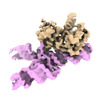 36046MC 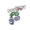 8hujC C: citing same article ( M: map data used to model this data |
|---|---|
| Similar structure data |
-Links
 PDBj
PDBj


- Assembly
| Deposited unit | 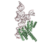
|
|---|---|
| 1 |
|
-Components
| #1: Protein | / eIF-4-gamma 1 / eIF-4G 1 / eIF-4G1 / p220 Mass: 31786.811 Da / Num. of mol.: 1 Source method: isolated from a genetically manipulated source Source: (gene. exp.) Homo sapiens (human) / Gene: EIF4G1, EIF4F, EIF4G, EIF4GI / Production host:  Escherichia coli (E. coli) / References: UniProt: Q04637 Escherichia coli (E. coli) / References: UniProt: Q04637 | ||||
|---|---|---|---|---|---|
| #2: RNA chain | Mass: 34838.609 Da / Num. of mol.: 1 / Source method: obtained synthetically / Source: (synth.) Encephalomyocarditis virus / References: GenBank: NC_001479.1 | ||||
| #3: Chemical |   Mass: 24.305 Da / Num. of mol.: 3 / Source method: obtained synthetically / Formula: Mg Mass: 24.305 Da / Num. of mol.: 3 / Source method: obtained synthetically / Formula: Mg#4: Water | ChemComp-HOH / | Water Mass: 18.015 Da / Num. of mol.: 1 / Source method: isolated from a natural source / Formula: H2O Mass: 18.015 Da / Num. of mol.: 1 / Source method: isolated from a natural source / Formula: H2OHas ligand of interest | N | |
-Experimental details
-Experiment
| Experiment | Method: ELECTRON MICROSCOPY |
|---|---|
| EM experiment | Aggregation state: PARTICLE / 3D reconstruction method: single particle reconstruction |
- Sample preparation
| Component | Name: Ternary complex of the J-K-St region of EMCV IRES with eIF4A and eIF4G-HEAT1 Type: COMPLEX / Entity ID: #1-#2 / Source: RECOMBINANT | ||||||||||||||||||||||||||||||
|---|---|---|---|---|---|---|---|---|---|---|---|---|---|---|---|---|---|---|---|---|---|---|---|---|---|---|---|---|---|---|---|
| Molecular weight |
| ||||||||||||||||||||||||||||||
| Source (natural) | Organism: Homo sapiens (human) | ||||||||||||||||||||||||||||||
| Source (recombinant) | Organism: Escherichia coli (E. coli) | ||||||||||||||||||||||||||||||
| Buffer solution | pH: 7.5 | ||||||||||||||||||||||||||||||
| Buffer component |
| ||||||||||||||||||||||||||||||
| Specimen | Embedding applied: NO / Shadowing applied: NO / Staining applied: NO / Vitrification applied: YES | ||||||||||||||||||||||||||||||
| Specimen support | Grid material: GOLD / Grid mesh size: 300 divisions/in. / Grid type: Quantifoil R1.2/1.3 | ||||||||||||||||||||||||||||||
| Vitrification | Instrument: FEI VITROBOT MARK IV / Cryogen name: ETHANE / Humidity: 95 % / Chamber temperature: 277 K |
- Electron microscopy imaging
| Microscopy | Model: JEOL CRYO ARM 300 Details: The microscope model is the JEOL's "JEM-Z320FHC", the custom-built model equipped with a helium-cooled specimen stage. |
|---|---|
| Electron gun | Electron source: FIELD EMISSION GUN / Accelerating voltage: 300 kV / Illumination mode: FLOOD BEAM |
| Electron lens | Mode: BRIGHT FIELDBright-field microscopy / Nominal defocus max: 2500 nm / Nominal defocus min: 1000 nm / Cs: 3.4 mm / Alignment procedure: COMA FREE |
| Specimen holder | Cryogen: NITROGEN |
| Image recording | Average exposure time: 8 sec. / Electron dose: 71.1 e/Å2 / Detector mode: COUNTING / Film or detector model: GATAN K2 SUMMIT (4k x 4k) / Num. of grids imaged: 2 / Num. of real images: 6194 |
| EM imaging optics | Energyfilter name: In-column Omega Filter / Energyfilter slit width: 20 eV |
| Image scans | Movie frames/image: 40 / Used frames/image: 1-40 |
- Processing
| Software | Name: PHENIX / Version: 1.20.1_4487: / Classification: refinement | ||||||||||||||||||||||||||||||||||||||||||||||||||||||||||||||||||||||||||||||||||||||||||||||||||||||||||
|---|---|---|---|---|---|---|---|---|---|---|---|---|---|---|---|---|---|---|---|---|---|---|---|---|---|---|---|---|---|---|---|---|---|---|---|---|---|---|---|---|---|---|---|---|---|---|---|---|---|---|---|---|---|---|---|---|---|---|---|---|---|---|---|---|---|---|---|---|---|---|---|---|---|---|---|---|---|---|---|---|---|---|---|---|---|---|---|---|---|---|---|---|---|---|---|---|---|---|---|---|---|---|---|---|---|---|---|
| EM software |
| ||||||||||||||||||||||||||||||||||||||||||||||||||||||||||||||||||||||||||||||||||||||||||||||||||||||||||
| CTF correction | Type: PHASE FLIPPING AND AMPLITUDE CORRECTION | ||||||||||||||||||||||||||||||||||||||||||||||||||||||||||||||||||||||||||||||||||||||||||||||||||||||||||
| Particle selection | Num. of particles selected: 947392 | ||||||||||||||||||||||||||||||||||||||||||||||||||||||||||||||||||||||||||||||||||||||||||||||||||||||||||
| Symmetry | Point symmetry: C1 (asymmetric) | ||||||||||||||||||||||||||||||||||||||||||||||||||||||||||||||||||||||||||||||||||||||||||||||||||||||||||
| 3D reconstruction | Resolution: 3.7 Å / Resolution method: FSC 0.143 CUT-OFF / Num. of particles: 255256 / Symmetry type: POINT | ||||||||||||||||||||||||||||||||||||||||||||||||||||||||||||||||||||||||||||||||||||||||||||||||||||||||||
| Atomic model building | Space: REAL | ||||||||||||||||||||||||||||||||||||||||||||||||||||||||||||||||||||||||||||||||||||||||||||||||||||||||||
| Refinement | Resolution: 3.7→107.86 Å / Cor.coef. Fo:Fc: 0.977 / SU B: 15.91 / SU ML: 0.235 / ESU R: 0.366 Stereochemistry target values: MAXIMUM LIKELIHOOD WITH PHASES Details: HYDROGENS HAVE BEEN ADDED IN THE RIDING POSITIONS
| ||||||||||||||||||||||||||||||||||||||||||||||||||||||||||||||||||||||||||||||||||||||||||||||||||||||||||
| Solvent computation | Solvent model: PARAMETERS FOR MASK CACLULATION | ||||||||||||||||||||||||||||||||||||||||||||||||||||||||||||||||||||||||||||||||||||||||||||||||||||||||||
| Displacement parameters | Biso mean: 199.643 Å2 | ||||||||||||||||||||||||||||||||||||||||||||||||||||||||||||||||||||||||||||||||||||||||||||||||||||||||||
| Refinement step | Cycle: 1 / Total: 4033 | ||||||||||||||||||||||||||||||||||||||||||||||||||||||||||||||||||||||||||||||||||||||||||||||||||||||||||
| Refine LS restraints |
|