 Movie
Movie Controller


+ Open data

- Basic information
| Entry | Database: PDB / ID: 8j2y | ||||||
|---|---|---|---|---|---|---|---|
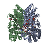
| Title | Acidimicrobiaceae bacterium photocobilins protein, dark state | ||||||
 Components Components | GGDEF domain-containing protein | ||||||
 Keywords Keywords | UNKNOWN FUNCTION /  cobalamin binding / biliverdin binding / B12-dependent photoreceptor protein / photocobilins cobalamin binding / biliverdin binding / B12-dependent photoreceptor protein / photocobilins | ||||||
| Function / homology |  Function and homology information Function and homology information | ||||||
| Biological species |  Acidimicrobiaceae bacterium (bacteria) Acidimicrobiaceae bacterium (bacteria) | ||||||
| Method | X-RAY DIFFRACTION / SYNCHROTRON / MOLECULAR REPLACEMENT / Resolution: 2.3 Å | ||||||
 Authors Authors | Zhang, S. / Poddar, H. / Levy, W.C. / Leys, D. | ||||||
| Funding support |  United Kingdom, 1items United Kingdom, 1items
| ||||||
 Citation Citation | Journal: Nat Commun / Year: 2024 Title: Photocobilins integrate B12 and bilin photochemistry for enzyme control. Authors: Zhang, S. / Jeffreys, L.N. / Poddar, H. / Yu, Y. / Liu, C. / Patel, K. / Johannissen, L.O. / Zhu, L. / Cliff, M.J. / Yan, C. / Schiro, G. / Weik, M. / Sakuma, M. / Levy, C.W. / Leys, D. / ...Authors: Zhang, S. / Jeffreys, L.N. / Poddar, H. / Yu, Y. / Liu, C. / Patel, K. / Johannissen, L.O. / Zhu, L. / Cliff, M.J. / Yan, C. / Schiro, G. / Weik, M. / Sakuma, M. / Levy, C.W. / Leys, D. / Heyes, D.J. / Scrutton, N.S. | ||||||
| History |
|
- Structure visualization
| Structure viewer | Molecule: MolmilJmol/JSmol |
|---|
- Downloads & links
-Download
| PDBx/mmCIF format | 8j2y.cif.gz | 277.3 KB | Display | PDBx/mmCIF format |
|---|---|---|---|---|
| PDB format | pdb8j2y.ent.gz | 223.9 KB | Display | PDB format |
| PDBx/mmJSON format | 8j2y.json.gz | Tree view | PDBx/mmJSON format | |
| Others |  Other downloads Other downloads |
-Validation report
| Arichive directory | https://data.pdbj.org/pub/pdb/validation_reports/j2/8j2yftp://data.pdbj.org/pub/pdb/validation_reports/j2/8j2y | HTTPS FTP |
|---|
-Related structure data

-Links
 PDBj
PDBj


- Assembly
| Deposited unit | 
| |||||||||
|---|---|---|---|---|---|---|---|---|---|---|
| 1 |
| |||||||||
| Unit cell |
| |||||||||
| Components on special symmetry positions |
|
-Components
-Protein , 1 types, 2 molecules AB
| #1: Protein | Mass: 37356.668 Da / Num. of mol.: 2 Source method: isolated from a genetically manipulated source Source: (gene. exp.) Acidimicrobiaceae bacterium (bacteria) / Gene: DCS55_04760 / Production host: Escherichia coli BL21(DE3) (bacteria) / References: UniProt: A0A349B205 |
|---|
-Non-polymers , 5 types, 135 molecules 


| #2: Chemical | Vitamin B12 Mass: 1330.356 Da / Num. of mol.: 2 / Source method: obtained synthetically / Formula: C62H89CoN13O14P / Feature type: SUBJECT OF INVESTIGATION Mass: 1330.356 Da / Num. of mol.: 2 / Source method: obtained synthetically / Formula: C62H89CoN13O14P / Feature type: SUBJECT OF INVESTIGATION#3: Chemical | Deoxyadenosine Mass: 251.242 Da / Num. of mol.: 2 / Source method: obtained synthetically / Formula: C10H13N5O3 / Feature type: SUBJECT OF INVESTIGATION Mass: 251.242 Da / Num. of mol.: 2 / Source method: obtained synthetically / Formula: C10H13N5O3 / Feature type: SUBJECT OF INVESTIGATION#4: Chemical | ChemComp-PEG / | Diethylene glycol Mass: 106.120 Da / Num. of mol.: 1 / Source method: obtained synthetically / Formula: C4H10O3 Mass: 106.120 Da / Num. of mol.: 1 / Source method: obtained synthetically / Formula: C4H10O3#5: Chemical | Thiocyanate Mass: 58.082 Da / Num. of mol.: 2 / Source method: obtained synthetically / Formula: CNS Mass: 58.082 Da / Num. of mol.: 2 / Source method: obtained synthetically / Formula: CNS#6: Water | ChemComp-HOH / | WaterMass: 18.015 Da / Num. of mol.: 128 / Source method: isolated from a natural source / Formula: H2O |
|---|
-Details
| Has ligand of interest | Y |
|---|
-Experimental details
-Experiment
| Experiment | Method: X-RAY DIFFRACTION / Number of used crystals: 1 |
|---|
- Sample preparation
| Crystal | Density Matthews: 2.85 Å3/Da / Density % sol: 56.9 % |
|---|---|
| Crystal grow | Temperature: 277 K / Method: vapor diffusion, sitting drop Details: 8% PEG 20,000, 8% PEG 550 MME, 0.1M Sodium acetate pH5.5, 0.2M Potassium thiocyanate |
-Data collection
| Diffraction | Mean temperature: 100 K / Serial crystal experiment: N |
|---|---|
| Diffraction source | Source: SYNCHROTRON / Site: Diamond / Beamline: I04 / Wavelength: 0.9795 Å |
| Detector | Type: DECTRIS EIGER2 XE 16M / Detector: PIXEL / Date: Oct 9, 2021 |
| Radiation | Protocol: SINGLE WAVELENGTH / Monochromatic (M) / Laue (L): M / Scattering type: x-ray |
| Radiation wavelength | Wavelength: 0.9795 Å / Relative weight: 1 |
| Reflection | Resolution: 2.3→44.31 Å / Num. obs: 38287 / % possible obs: 99.86 % / Redundancy: 13.7 % / Rmerge(I) obs: 0.1766 / Net I/σ(I): 6.95 |
| Reflection shell | Resolution: 2.3→2.382 Å / Redundancy: 13.3 % / Rmerge(I) obs: 0.9439 / Mean I/σ(I) obs: 0.94 / Num. unique obs: 3775 / % possible all: 99.79 |
- Processing
| Software |
| |||||||||||||||||||||||||||||||||||||||||||||||||||||||||||||||||||||||||||||||||||||||||||||||
|---|---|---|---|---|---|---|---|---|---|---|---|---|---|---|---|---|---|---|---|---|---|---|---|---|---|---|---|---|---|---|---|---|---|---|---|---|---|---|---|---|---|---|---|---|---|---|---|---|---|---|---|---|---|---|---|---|---|---|---|---|---|---|---|---|---|---|---|---|---|---|---|---|---|---|---|---|---|---|---|---|---|---|---|---|---|---|---|---|---|---|---|---|---|---|---|---|
| Refinement | Method to determine structure: MOLECULAR REPLACEMENT / Resolution: 2.3→44.31 Å / Cor.coef. Fo:Fc: 0.952 / Cor.coef. Fo:Fc free: 0.947 / SU B: 20.306 / SU ML: 0.231 / Cross valid method: THROUGHOUT / ESU R: 0.288 / ESU R Free: 0.211 / Stereochemistry target values: MAXIMUM LIKELIHOOD Details: U VALUES : WITH TLS ADDED HYDROGENS HAVE BEEN ADDED IN THE RIDING POSITIONS U VALUES : RESIDUAL ONLY
| |||||||||||||||||||||||||||||||||||||||||||||||||||||||||||||||||||||||||||||||||||||||||||||||
| Solvent computation | Ion probe radii: 0.8 Å / Shrinkage radii: 0.8 Å / VDW probe radii: 1.1 Å / Solvent model: MASK | |||||||||||||||||||||||||||||||||||||||||||||||||||||||||||||||||||||||||||||||||||||||||||||||
| Displacement parameters | Biso mean: 63.721 Å2
| |||||||||||||||||||||||||||||||||||||||||||||||||||||||||||||||||||||||||||||||||||||||||||||||
| Refinement step | Cycle: LAST / Resolution: 2.3→44.31 Å
| |||||||||||||||||||||||||||||||||||||||||||||||||||||||||||||||||||||||||||||||||||||||||||||||
| Refine LS restraints |
| |||||||||||||||||||||||||||||||||||||||||||||||||||||||||||||||||||||||||||||||||||||||||||||||
| LS refinement shell | Resolution: 2.3→2.359 Å / Total num. of bins used: 20
| |||||||||||||||||||||||||||||||||||||||||||||||||||||||||||||||||||||||||||||||||||||||||||||||
| Refinement TLS params. | Method: refined / Refine-ID: X-RAY DIFFRACTION
| |||||||||||||||||||||||||||||||||||||||||||||||||||||||||||||||||||||||||||||||||||||||||||||||
| Refinement TLS group |
|
