 Movie
Movie Controller

[English] 日本語
 Yorodumi
- PDB-8iyo: Crystal structure of a protein acetyltransferase, HP0935, acetyl-... -
+ Open data

- Basic information
| Entry | Database: PDB / ID: 8iyo | ||||||
|---|---|---|---|---|---|---|---|
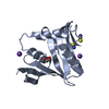
| Title | Crystal structure of a protein acetyltransferase, HP0935, acetyl-CoA bound form | ||||||
 Components Components | N-acetyltransferase domain-containing protein | ||||||
 Keywords Keywords |  TRANSFERASE / GNAT domain / protein acetyltransferase / acetyl-coenzyme A TRANSFERASE / GNAT domain / protein acetyltransferase / acetyl-coenzyme A | ||||||
| Function / homology | acyltransferase activity, transferring groups other than amino-acyl groups / Acetyltransferase (GNAT) family / Gcn5-related N-acetyltransferase (GNAT) domain profile. / GNAT domain / Acyl-CoA N-acyltransferase / ACETYL COENZYME *A / N-acetyltransferase domain-containing protein Function and homology information Function and homology information | ||||||
| Biological species |  Helicobacter pylori 26695 (bacteria) Helicobacter pylori 26695 (bacteria) | ||||||
| Method | X-RAY DIFFRACTION / SYNCHROTRON / MOLECULAR REPLACEMENT / molecular replacement / Resolution: 2.4 Å | ||||||
 Authors Authors | Dadireddy, V. / Mahanta, P. / Kumar, A. / Desirazu, R.N. / Ramakumar, S. | ||||||
| Funding support | 1items
| ||||||
 Citation Citation | Journal: To be published Title: Crystal structure of a protein acetyltransferase, HP0935, acetyl-CoA bound form Authors: Dadireddy, V. / Mahanta, P. / Kumar, A. / Desirazu, R.N. / Ramakumar, S. | ||||||
| History |
|
- Structure visualization
| Structure viewer | Molecule: MolmilJmol/JSmol |
|---|
- Downloads & links
-Download
| PDBx/mmCIF format | 8iyo.cif.gz | 277.7 KB | Display | PDBx/mmCIF format |
|---|---|---|---|---|
| PDB format | pdb8iyo.ent.gz | 227.7 KB | Display | PDB format |
| PDBx/mmJSON format | 8iyo.json.gz | Tree view | PDBx/mmJSON format | |
| Others |  Other downloads Other downloads |
-Validation report
| Arichive directory | https://data.pdbj.org/pub/pdb/validation_reports/iy/8iyoftp://data.pdbj.org/pub/pdb/validation_reports/iy/8iyo | HTTPS FTP |
|---|
-Related structure data
| Related structure data | |
|---|---|
| Similar structure data |
-Links
 PDBj
PDBj

- Assembly
| Deposited unit | 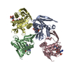
| ||||||||
|---|---|---|---|---|---|---|---|---|---|
| 1 | 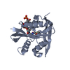
| ||||||||
| 2 | 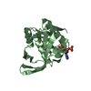
| ||||||||
| 3 | 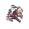
| ||||||||
| 4 | 
| ||||||||
| Unit cell |
|
-Components
| #1: Protein | Mass: 18626.504 Da / Num. of mol.: 4 / Mutation: M1V Source method: isolated from a genetically manipulated source Source: (gene. exp.) Helicobacter pylori 26695 (bacteria) / Gene: HP_0935 / Plasmid: pNIC-Bsa4 / Production host: Escherichia coli BL21(DE3) (bacteria) / Variant (production host): star / References: UniProt: O25589#2: Chemical | ChemComp-ACO / Acetyl-CoA  Mass: 809.571 Da / Num. of mol.: 4 / Source method: obtained synthetically / Formula: C23H38N7O17P3S / Feature type: SUBJECT OF INVESTIGATION Mass: 809.571 Da / Num. of mol.: 4 / Source method: obtained synthetically / Formula: C23H38N7O17P3S / Feature type: SUBJECT OF INVESTIGATION#3: Water | ChemComp-HOH / | Water Mass: 18.015 Da / Num. of mol.: 75 / Source method: isolated from a natural source / Formula: H2O Mass: 18.015 Da / Num. of mol.: 75 / Source method: isolated from a natural source / Formula: H2OHas ligand of interest | Y | |
|---|
-Experimental details
-Experiment
| Experiment | Method: X-RAY DIFFRACTION / Number of used crystals: 1 |
|---|
- Sample preparation
| Crystal | Density Matthews: 3.23 Å3/Da / Density % sol: 61.93 % / Description: Needles |
|---|---|
| Crystal grow | Temperature: 277 K / Method: vapor diffusion, sitting drop / pH: 7.5 Details: 0.02M magnesium chloride hexahydrate, 0.1M HEPES pH 7.5, 22% (w/v) poly(acrylic acid, sodium salt) 5100, ethylene glycol (cryo, soaked) |
-Data collection
| Diffraction | Mean temperature: 100 K / Serial crystal experiment: N | ||||||||||||||||||||||||||||||
|---|---|---|---|---|---|---|---|---|---|---|---|---|---|---|---|---|---|---|---|---|---|---|---|---|---|---|---|---|---|---|---|
| Diffraction source | Source: SYNCHROTRON / Site: ESRF  / Beamline: ID29 / Wavelength: 1.07227 Å / Beamline: ID29 / Wavelength: 1.07227 Å | ||||||||||||||||||||||||||||||
| Detector | Type: DECTRIS PILATUS3 6M / Detector: PIXEL / Date: Dec 2, 2018 | ||||||||||||||||||||||||||||||
| Radiation | Protocol: SINGLE WAVELENGTH / Monochromatic (M) / Laue (L): M / Scattering type: x-ray | ||||||||||||||||||||||||||||||
| Radiation wavelength | Wavelength: 1.07227 Å / Relative weight: 1 | ||||||||||||||||||||||||||||||
| Reflection | Resolution: 2.4→44.08 Å / Num. obs: 36841 / % possible obs: 100 % / Redundancy: 18.3 % / Biso Wilson estimate: 46.76 Å2 / CC1/2: 1 / Rmerge(I) obs: 0.15 / Rpim(I) all: 0.036 / Rrim(I) all: 0.154 / Net I/σ(I): 15.2 / Num. measured all: 675020 / Scaling rejects: 3 | ||||||||||||||||||||||||||||||
| Reflection shell | Diffraction-ID: 1
|
-Phasing
| Phasing | Method: molecular replacement | |||||||||
|---|---|---|---|---|---|---|---|---|---|---|
| Phasing MR | Model details: Phaser MODE: MR_AUTO
|
- Processing
| Software |
| |||||||||||||||||||||||||||||||||||||||||||||||||||||||||||||||||||||||||||||||||||||||||||||||||||||||||||||||||||||||||||||
|---|---|---|---|---|---|---|---|---|---|---|---|---|---|---|---|---|---|---|---|---|---|---|---|---|---|---|---|---|---|---|---|---|---|---|---|---|---|---|---|---|---|---|---|---|---|---|---|---|---|---|---|---|---|---|---|---|---|---|---|---|---|---|---|---|---|---|---|---|---|---|---|---|---|---|---|---|---|---|---|---|---|---|---|---|---|---|---|---|---|---|---|---|---|---|---|---|---|---|---|---|---|---|---|---|---|---|---|---|---|---|---|---|---|---|---|---|---|---|---|---|---|---|---|---|---|---|
| Refinement | Method to determine structure: MOLECULAR REPLACEMENT / Resolution: 2.4→44.08 Å / SU ML: 0.29 / Cross valid method: THROUGHOUT / σ(F): 1.34 / Phase error: 25.14 / Stereochemistry target values: ML
| |||||||||||||||||||||||||||||||||||||||||||||||||||||||||||||||||||||||||||||||||||||||||||||||||||||||||||||||||||||||||||||
| Solvent computation | Shrinkage radii: 0.9 Å / VDW probe radii: 1.1 Å / Solvent model: FLAT BULK SOLVENT MODEL | |||||||||||||||||||||||||||||||||||||||||||||||||||||||||||||||||||||||||||||||||||||||||||||||||||||||||||||||||||||||||||||
| Displacement parameters | Biso max: 123.77 Å2 / Biso mean: 58.7408 Å2 / Biso min: 33.12 Å2 | |||||||||||||||||||||||||||||||||||||||||||||||||||||||||||||||||||||||||||||||||||||||||||||||||||||||||||||||||||||||||||||
| Refinement step | Cycle: final / Resolution: 2.4→44.08 Å
| |||||||||||||||||||||||||||||||||||||||||||||||||||||||||||||||||||||||||||||||||||||||||||||||||||||||||||||||||||||||||||||
| LS refinement shell | Refine-ID: X-RAY DIFFRACTION / Rfactor Rfree error: 0 / Total num. of bins used: 13 / % reflection obs: 100 %
| |||||||||||||||||||||||||||||||||||||||||||||||||||||||||||||||||||||||||||||||||||||||||||||||||||||||||||||||||||||||||||||
| Refinement TLS params. | Method: refined / Refine-ID: X-RAY DIFFRACTION
| |||||||||||||||||||||||||||||||||||||||||||||||||||||||||||||||||||||||||||||||||||||||||||||||||||||||||||||||||||||||||||||
| Refinement TLS group |
|
