 Movie
Movie Controller

[English] 日本語
 Yorodumi
- PDB-8iua: Crystal structure of GH66 endodextranase from Flavobacterium john... -
+ Open data

- Basic information
| Entry | Database: PDB / ID: 8iua | ||||||
|---|---|---|---|---|---|---|---|
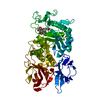
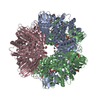
| Title | Crystal structure of GH66 endodextranase from Flavobacterium johnsoniae in complex with isomaltose | ||||||
 Components Components | Candidate dextranase Glycoside hydrolase family 66 | ||||||
 Keywords Keywords |  HYDROLASE / glycoside hydrolase / GH66 / dextran / TIM-barrel HYDROLASE / glycoside hydrolase / GH66 / dextran / TIM-barrel | ||||||
| Function / homology | Glycosyl hydrolase family 66 / Glycosyl hydrolase family 66 / Glycosyl hydrolase, all-beta / Glycoside hydrolase superfamily / hydrolase activity / Immunoglobulin-like fold / beta-D-glucopyranose / Candidate dextranase Glycoside hydrolase family 66 Function and homology information Function and homology information | ||||||
| Biological species |  Flavobacterium johnsoniae UW101 (bacteria) Flavobacterium johnsoniae UW101 (bacteria) | ||||||
| Method | X-RAY DIFFRACTION / SYNCHROTRON / MOLECULAR REPLACEMENT / Resolution: 1.8 Å | ||||||
 Authors Authors | Nakamura, S. / Miyazaki, T. | ||||||
| Funding support |  Japan, 1items Japan, 1items
| ||||||
 Citation Citation | Journal: J.Biol.Chem. / Year: 2023 Title: Bacteroidota polysaccharide utilization system for branched dextran exopolysaccharides from lactic acid bacteria. Authors: Nakamura, S. / Kurata, R. / Tonozuka, T. / Funane, K. / Park, E.Y. / Miyazaki, T. | ||||||
| History |
|
- Structure visualization
| Structure viewer | Molecule: MolmilJmol/JSmol |
|---|
- Downloads & links
-Download
| PDBx/mmCIF format | 8iua.cif.gz | 546.2 KB | Display | PDBx/mmCIF format |
|---|---|---|---|---|
| PDB format | pdb8iua.ent.gz | 345.3 KB | Display | PDB format |
| PDBx/mmJSON format | 8iua.json.gz | Tree view | PDBx/mmJSON format | |
| Others |  Other downloads Other downloads |
-Validation report
| Arichive directory | https://data.pdbj.org/pub/pdb/validation_reports/iu/8iuaftp://data.pdbj.org/pub/pdb/validation_reports/iu/8iua | HTTPS FTP |
|---|
-Related structure data


-Links
 PDBj
PDBj


- Assembly
| Deposited unit | 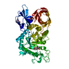
| ||||||||
|---|---|---|---|---|---|---|---|---|---|
| 1 |
| ||||||||
| Unit cell |
| ||||||||
| Components on special symmetry positions |
|
-Components
| #1: Protein | Mass: 64578.754 Da / Num. of mol.: 1 Source method: isolated from a genetically manipulated source Source: (gene. exp.) Flavobacterium johnsoniae UW101 (bacteria)Gene: Fjoh_4431 / Plasmid: pET28a / Production host: Escherichia coli BL21(DE3) (bacteria) / References: UniProt: A5FBI2 |
|---|---|
| #2: Polysaccharide | alpha-D-glucopyranose-(1-6)-alpha-D-glucopyranose / Mass: 342.297 Da / Num. of mol.: 1 Source method: isolated from a genetically manipulated source |
| #3: Sugar | ChemComp-BGC / Glucose  Type: D-saccharide, beta linking / Mass: 180.156 Da / Num. of mol.: 1 / Source method: obtained synthetically / Formula: C6H12O6 / Feature type: SUBJECT OF INVESTIGATION Type: D-saccharide, beta linking / Mass: 180.156 Da / Num. of mol.: 1 / Source method: obtained synthetically / Formula: C6H12O6 / Feature type: SUBJECT OF INVESTIGATION |
| #4: Chemical | ChemComp-NA /   Mass: 22.990 Da / Num. of mol.: 1 / Source method: obtained synthetically / Formula: Na Mass: 22.990 Da / Num. of mol.: 1 / Source method: obtained synthetically / Formula: Na |
| #5: Water | ChemComp-HOH / Water Mass: 18.015 Da / Num. of mol.: 268 / Source method: isolated from a natural source / Formula: H2O Mass: 18.015 Da / Num. of mol.: 268 / Source method: isolated from a natural source / Formula: H2O |
| Has ligand of interest | Y |
-Experimental details
-Experiment
| Experiment | Method: X-RAY DIFFRACTION / Number of used crystals: 1 |
|---|
- Sample preparation
| Crystal | Density Matthews: 2.11 Å3/Da / Density % sol: 41.8 % |
|---|---|
| Crystal grow | Temperature: 293 K / Method: vapor diffusion, hanging drop Details: 100mM Tris-HCl, pH 8.5-9.0, 200mM lithium sulfate, 20% PEG 4000 PH range: 8.5-9.0 |
-Data collection
| Diffraction | Mean temperature: 100 K / Serial crystal experiment: N |
|---|---|
| Diffraction source | Source: SYNCHROTRON / Site: Photon Factory / Beamline: BL-5A / Wavelength: 1 Å |
| Detector | Type: DECTRIS PILATUS 2M / Detector: PIXEL / Date: Mar 5, 2022 |
| Radiation | Protocol: SINGLE WAVELENGTH / Monochromatic (M) / Laue (L): M / Scattering type: x-ray |
| Radiation wavelength | Wavelength: 1 Å / Relative weight: 1 |
| Reflection | Resolution: 1.8→46.9 Å / Num. obs: 50250 / % possible obs: 99.7 % / Redundancy: 6.7 % / CC1/2: 0.997 / Rmerge(I) obs: 0.121 / Rpim(I) all: 0.077 / Rrim(I) all: 0.144 / Net I/σ(I): 11 |
| Reflection shell | Resolution: 1.8→1.84 Å / Rmerge(I) obs: 0.92 / Mean I/σ(I) obs: 2.3 / Num. unique obs: 50250 / CC1/2: 0.839 / Rpim(I) all: 0.57 / Rrim(I) all: 1.085 |
- Processing
| Software |
| ||||||||||||||||||||||||||||||||||||||||||||||||||||||||||||||||||||||||||||||||||||||||||||||||||||||||||||||||||||||||||||||||||||||||||||||||||||||||||||||||
|---|---|---|---|---|---|---|---|---|---|---|---|---|---|---|---|---|---|---|---|---|---|---|---|---|---|---|---|---|---|---|---|---|---|---|---|---|---|---|---|---|---|---|---|---|---|---|---|---|---|---|---|---|---|---|---|---|---|---|---|---|---|---|---|---|---|---|---|---|---|---|---|---|---|---|---|---|---|---|---|---|---|---|---|---|---|---|---|---|---|---|---|---|---|---|---|---|---|---|---|---|---|---|---|---|---|---|---|---|---|---|---|---|---|---|---|---|---|---|---|---|---|---|---|---|---|---|---|---|---|---|---|---|---|---|---|---|---|---|---|---|---|---|---|---|---|---|---|---|---|---|---|---|---|---|---|---|---|---|---|---|---|
| Refinement | Method to determine structure: MOLECULAR REPLACEMENT / Resolution: 1.8→46.9 Å / Cor.coef. Fo:Fc: 0.953 / Cor.coef. Fo:Fc free: 0.927 / SU B: 10.231 / SU ML: 0.153 / Cross valid method: FREE R-VALUE / ESU R: 0.154 / ESU R Free: 0.145 Details: Hydrogens have been added in their riding positions
| ||||||||||||||||||||||||||||||||||||||||||||||||||||||||||||||||||||||||||||||||||||||||||||||||||||||||||||||||||||||||||||||||||||||||||||||||||||||||||||||||
| Solvent computation | Ion probe radii: 0.8 Å / Shrinkage radii: 0.8 Å / VDW probe radii: 1.2 Å / Solvent model: MASK BULK SOLVENT | ||||||||||||||||||||||||||||||||||||||||||||||||||||||||||||||||||||||||||||||||||||||||||||||||||||||||||||||||||||||||||||||||||||||||||||||||||||||||||||||||
| Displacement parameters | Biso mean: 28.622 Å2
| ||||||||||||||||||||||||||||||||||||||||||||||||||||||||||||||||||||||||||||||||||||||||||||||||||||||||||||||||||||||||||||||||||||||||||||||||||||||||||||||||
| Refinement step | Cycle: LAST / Resolution: 1.8→46.9 Å
| ||||||||||||||||||||||||||||||||||||||||||||||||||||||||||||||||||||||||||||||||||||||||||||||||||||||||||||||||||||||||||||||||||||||||||||||||||||||||||||||||
| Refine LS restraints |
| ||||||||||||||||||||||||||||||||||||||||||||||||||||||||||||||||||||||||||||||||||||||||||||||||||||||||||||||||||||||||||||||||||||||||||||||||||||||||||||||||
| LS refinement shell |
| ||||||||||||||||||||||||||||||||||||||||||||||||||||||||||||||||||||||||||||||||||||||||||||||||||||||||||||||||||||||||||||||||||||||||||||||||||||||||||||||||
| Refinement TLS params. | Method: refined / Refine-ID: X-RAY DIFFRACTION
| ||||||||||||||||||||||||||||||||||||||||||||||||||||||||||||||||||||||||||||||||||||||||||||||||||||||||||||||||||||||||||||||||||||||||||||||||||||||||||||||||
| Refinement TLS group |
|
